Ameloblastoma CT: Difference between revisions
(Category) |
|||
| Line 3: | Line 3: | ||
{{CMG}}{{AE}}{{Simrat}} | {{CMG}}{{AE}}{{Simrat}} | ||
==Overview== | ==Overview== | ||
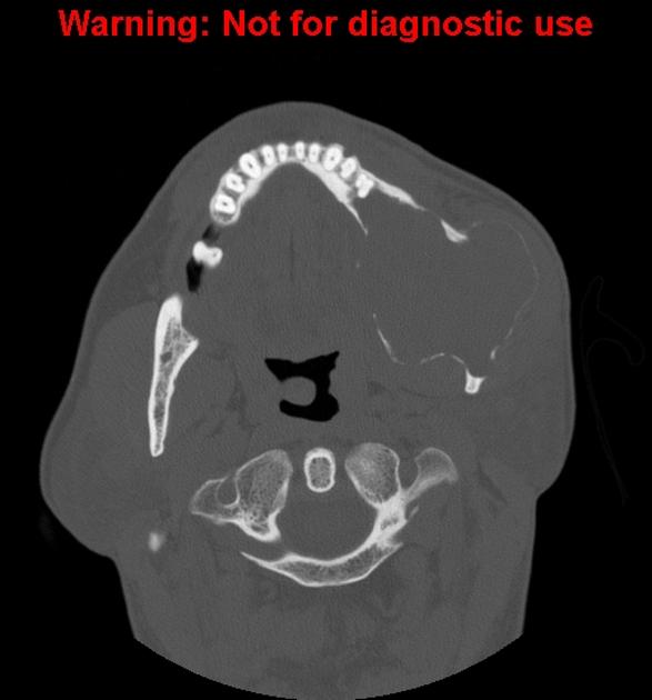
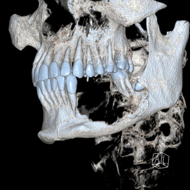
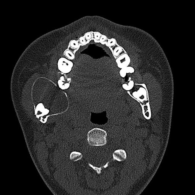
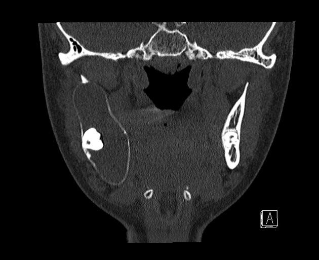
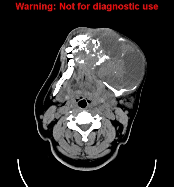
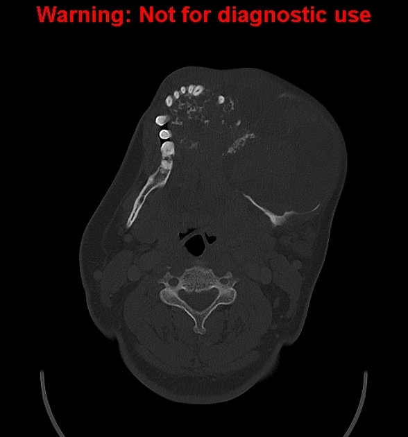
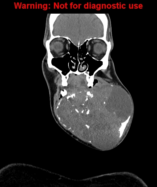
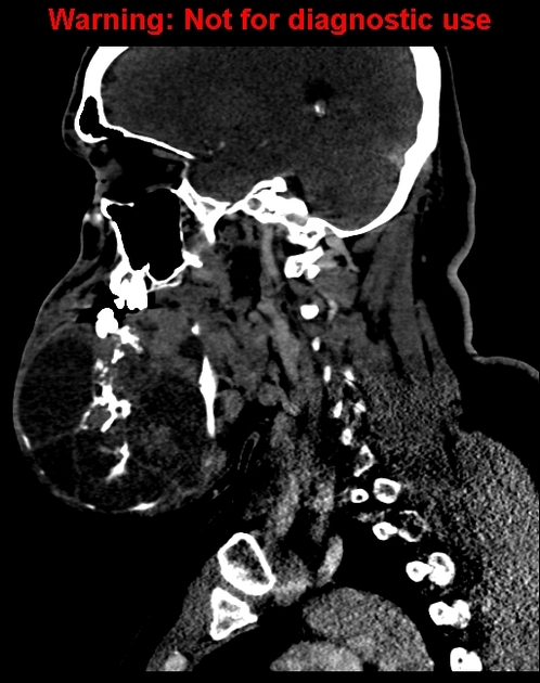
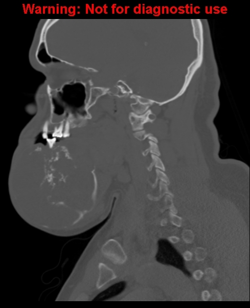
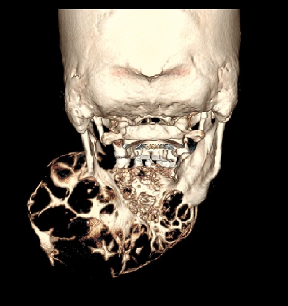
On head and neck CT, ameloblastoma is characterized by multiloculated, expansile "soap-bubble" lesion, with well demarcated borders, no matrix calcification, and occasionally erosion of the adjacent tooth roots. | On head and neck CT, ameloblastoma is characterized by multiloculated, expansile "soap-bubble" lesion, with well demarcated borders, no matrix calcification, and occasionally erosion of the adjacent tooth roots. | ||
==CT== | ==CT== | ||
Head and neck CT is classically seen as a multiloculated (80%), expansile "soap-bubble" lesion, with well demarcated borders and no matrix calcification. Occasionally erosion of the adjacent tooth roots can be seen which is highly specific. When larger it may also erode through cortex into adjacent soft tissues. | Head and neck CT is classically seen as a multiloculated (80%), expansile "soap-bubble" lesion, with well demarcated borders and no matrix calcification. Occasionally erosion of the adjacent tooth roots can be seen which is highly specific. When larger it may also erode through cortex into adjacent soft tissues. | ||
<gallery> | <gallery> | ||
Ameloblastoma CT.jpg|Ameloblastoma CT | File:Ameloblastoma CT.jpg|Ameloblastoma CT | ||
Axial bone marrow ameloblastoma 1.jpg|Axial bone window | File:Axial bone marrow ameloblastoma 1.jpg|Axial bone window | ||
Axial liver window ameloblastoma.jpg|Axial liver window ameloblastoma | File:Axial liver window ameloblastoma.jpg|Axial liver window ameloblastoma | ||
Coronal bone window ameloblastoma.jpg|Coronal bone window ameloblastoma | File:Coronal bone window ameloblastoma.jpg|Coronal bone window ameloblastoma | ||
Coronal liver window ameloblastoma.jpg|Coronal bone window ameloblastoma | File:Coronal liver window ameloblastoma.jpg|Coronal bone window ameloblastoma | ||
Axial non contrast ameloblastoma.jpg|Axial non contrast ameloblastoma | File:Axial non contrast ameloblastoma.jpg|Axial non contrast ameloblastoma | ||
Coronal non contrast ameloblastoma x ray.jpg|Coronal non contrast ameloblastoma<ref name="radio1">Image courtesy of Dr. | File:Coronal non contrast ameloblastoma x ray.jpg|Coronal non contrast ameloblastoma<ref name="radio1">Image courtesy of Dr. Frank Gaillard [http://www.radiopaedia.org Radiopaedia] (original file [http://radiopaedia.org/cases/ameloblastoma]).[http://radiopaedia.org/licence Creative Commons BY-SA-NC</ref> | ||
3D rendered volume.jpg|3 D volume rendered ameloblastoma | File:3D rendered volume.jpg|3 D volume rendered ameloblastoma | ||
3 d ameloblastoma.jpg|3 d ameloblastoma | File:3 d ameloblastoma.jpg|3 d ameloblastoma | ||
3D ameloblastoma.jpg|3D ameloblastoma | File:3D ameloblastoma.jpg|3D ameloblastoma | ||
Bone window 1 ameloblastoma.jpg|bone window ameloblastoma | File:Bone window 1 ameloblastoma.jpg|bone window ameloblastoma | ||
Bone window ameloblastoma.jpg|Bone Window ameloblastoma | File:Bone window ameloblastoma.jpg|Bone Window ameloblastoma | ||
Axial C- soft tissue window.jpg|Bone Window ameloblastoma | File:Axial C- soft tissue window.jpg|Bone Window ameloblastoma | ||
Axial bone window ameloblastoma.jpg|Axial Bone Window ameloblastoma | File:Axial bone window ameloblastoma.jpg|Axial Bone Window ameloblastoma | ||
Coronal soft tissue window ameloblastoma.jpg|coronal soft tissue Window ameloblastoma | File:Coronal soft tissue window ameloblastoma.jpg|coronal soft tissue Window ameloblastoma | ||
Coronal bone window ameloblastoma.jpg|coronal soft tissue Window ameloblastoma | File:Coronal bone window ameloblastoma.jpg|coronal soft tissue Window ameloblastoma | ||
Saggital soft tissue window ameloblastoma.jpg|saggital soft tissue Window ameloblastoma | File:Saggital soft tissue window ameloblastoma.jpg|saggital soft tissue Window ameloblastoma | ||
Saggital bone window ameloblastoma.jpg|saggital bone Window ameloblastoma | File:Saggital bone window ameloblastoma.jpg|saggital bone Window ameloblastoma | ||
3 d amelo.jpg|3 D ameloblastoma | File:3 d amelo.jpg|3 D ameloblastoma | ||
</gallery> | </gallery> | ||
==References== | ==References== | ||
Revision as of 14:23, 10 October 2018
|
Ameloblastoma Microchapters |
|
Diagnosis |
|---|
|
Treatment |
|
Case Studies |
|
Ameloblastoma CT On the Web |
|
American Roentgen Ray Society Images of Ameloblastoma CT |
Editor-In-Chief: C. Michael Gibson, M.S., M.D. [2]Associate Editor(s)-in-Chief: Simrat Sarai, M.D. [3]
Overview
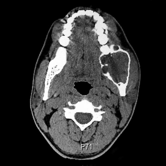
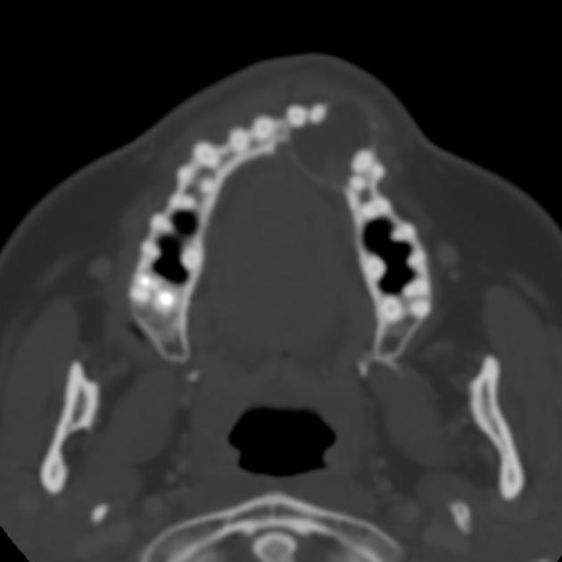
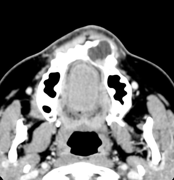
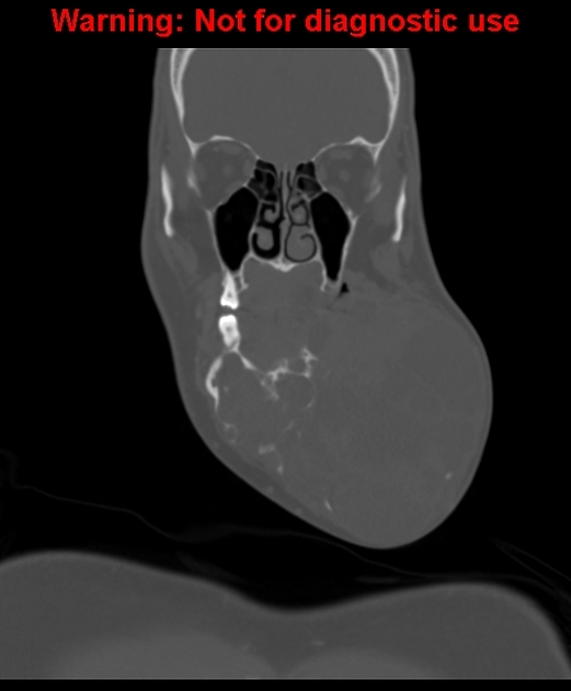
On head and neck CT, ameloblastoma is characterized by multiloculated, expansile "soap-bubble" lesion, with well demarcated borders, no matrix calcification, and occasionally erosion of the adjacent tooth roots.
CT
Head and neck CT is classically seen as a multiloculated (80%), expansile "soap-bubble" lesion, with well demarcated borders and no matrix calcification. Occasionally erosion of the adjacent tooth roots can be seen which is highly specific. When larger it may also erode through cortex into adjacent soft tissues.
-
Ameloblastoma CT
-
Axial bone window
-
Axial liver window ameloblastoma
-
Coronal bone window ameloblastoma
-
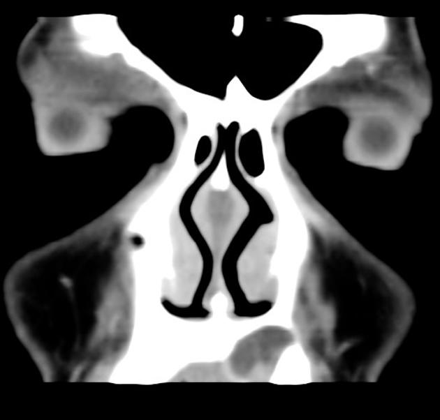
Coronal bone window ameloblastoma
-
Axial non contrast ameloblastoma
-
![Coronal non contrast ameloblastoma[1]](/images/6/63/Coronal_non_contrast_ameloblastoma_x_ray.jpg)
Coronal non contrast ameloblastoma[1]
-
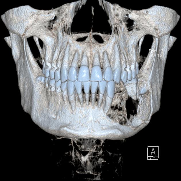
3 D volume rendered ameloblastoma
-
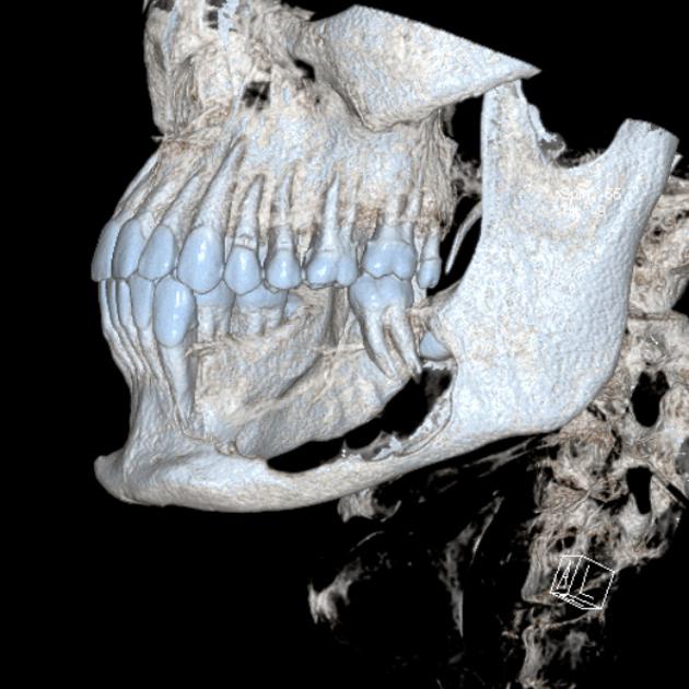
3 d ameloblastoma
-
3D ameloblastoma
-
bone window ameloblastoma
-
Bone Window ameloblastoma
-
Bone Window ameloblastoma
-
Axial Bone Window ameloblastoma
-
coronal soft tissue Window ameloblastoma
-
coronal soft tissue Window ameloblastoma
-
saggital soft tissue Window ameloblastoma
-
saggital bone Window ameloblastoma
-
3 D ameloblastoma


![Coronal non contrast ameloblastoma[1]](/index.php/File:Coronal_non_contrast_ameloblastoma_x_ray.jpg)


References
- ↑ Image courtesy of Dr. Frank Gaillard Radiopaedia (original file [1]).[http://radiopaedia.org/licence Creative Commons BY-SA-NC