Cystic medial necrosis: Difference between revisions
| Line 66: | Line 66: | ||
{{Reflist|2}} | {{Reflist|2}} | ||
[[Category:Disease]] | [[Category:Disease]] | ||
Latest revision as of 14:41, 11 August 2013
| Cystic medial necrosis | |
 | |
|---|---|
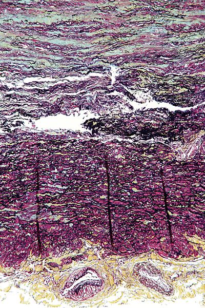
| Micrograph showing cystic medial degeneration, the histologic correlate of familial thoracic aortic aneurysms. The image shows abundant basophilic ground substance in the tunica media (blue at top of image) and disruption of the elastic fibers. The tunica adventitia (yellow at bottom of image) with vaso vasorum is also seen. Movat's stain. | |
| ICD-9 | 441.00 |
| OMIM | 607086 |
| DiseasesDB | 30073 |
|
Aortic aneurysm Microchapters |
|
Diagnosis |
|---|
|
Treatment |
|
Cystic medial necrosis On the Web |
|
Risk calculators and risk factors for Cystic medial necrosis |
Editor-In-Chief: C. Michael Gibson, M.S., M.D. [1]
Synonyms and keywords: cystic medial degeneration; familial thoracic aortic aneurysm and dissection; FTAAD; familial thoracic aortic aneurysm; familial aortic dissection; cystic medial necrosis of aorta [1]; it is sometimes called "Erdheim's cystic medial necrosis", after Jakob Erdheim[2][3]; the term cystic medial degeneration is sometimes used instead of cystic medial necrosis, because necrosis is not always found; Erdheim disease, medionecrosis aortae idiopathica cystica, medionecrosis of the aorta, mucoid medial degeneration
Overview
Cystic medial necrosis is an autosomal dominant[1] disorder of large arteries. There is a degenerative breakdown of collagen, elastin, and smooth muscle caused by aging which contributes to the weakening of the wall of the aorta. This weakening of the wall of the aorta can result in the formation of an aneurysm.
Pathology
Cystic media necrosis is a degenerative breakdown of collagen, elastin, and smooth muscle caused by aging which contributes to weakening of the wall of the artery. There is a loss of elastic and muscle fibers in the medial layer of the aorta along with the accumulation of mucopolysaccharides in cystlike spaces between the fibers (hence the term cystic). In the aorta, this can result in the formation of a fusiform aneurysm, and a subsequent risk of aortic dissection.
Genetics
Genetic variants include ACTA2 as well as:
| Type | OMIM | Gene | Locus |
|---|---|---|---|
| AAT1 | 607086 | 11q23.3-q24 | |
| AAT4 | 132900 | MYH11 | 16p |
| AAT6 | 611788 | ACTA2 | 10q |
Associated Conditions
There is an association between cystic medial necrosis and the following disorders:
Epidemiology and Demographics
The disease tends to percent after 40 years of age and is twice as common in men as in women.
Natural History, Complications, and Prognosis
Cystic medial necrosis is associated with an increased risk of aortic dissection.
References
- ↑ 1.0 1.1 Online Mendelian Inheritance in Man (OMIM) 607086
- ↑ Template:WhoNamedIt
- ↑ J. Erdheim. Medionecrosis aortae idiopathica (cystica). Archiv für pathologische Anatomie und Physiologie und für klinische Medizin, 1929, 273: 454-479.
- ↑ Myxomatous Degeneration and Cystic Medial Necrosis Associated With Acromegaly Sharon M. Ondreyco, MD; H. Daniel Lewis, Jr, MD; Charles R. Hartman, MD Arch Intern Med. 1980;140(4):547-549. doi:10.1001/archinte.1980.00330160107039