Solitary pulmonary nodule other imaging findings: Difference between revisions
Jump to navigation
Jump to search
(Created page with "__NOTOC__ {{Solitary pulmonary nodule}} {{CMG}} ==Overview== ==PET Scan== <div align="left"> <gallery heights="200" widths="200"> Image:Pulmonary AVM as nodule 1.jpg|Chest x-...") |
(No difference)
|
Revision as of 14:29, 25 September 2012
|
Pulmonary Nodule Microchapters |
|
Diagnosis |
|---|
|
Treatment |
|
Case Studies |
|
Solitary pulmonary nodule other imaging findings On the Web |
|
American Roentgen Ray Society Images of Solitary pulmonary nodule other imaging findings |
|
Solitary pulmonary nodule other imaging findings in the news |
|
Risk calculators and risk factors for Solitary pulmonary nodule other imaging findings |
Editor-In-Chief: C. Michael Gibson, M.S., M.D. [1]
Overview
PET Scan
-
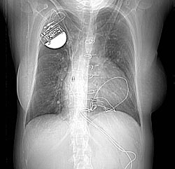
Chest x-ray: A 32 year old woman. 1. Two pulmonary arteriovenous malformations consistent with the nodules seen on the recent chest film. There is breathing artifact on several of the images and other tiny AVMs cannot be excluded. 2. Cardiomegaly with right atrial and left atrial enlargement and hepatic congestion.
-

Thorax CT

