File:VZV07.jpeg: Difference between revisions
Jump to navigation
Jump to search
(Under a relatively low magnification of 50x, this hematoxylin-eosin (H&E)-stained photomicrograph reveals some of the cytoarchitectural histopathologic changes which you’d find in a human skin tissue specimen that included a chickenpox, or varicella ...) |
(No difference)
|
Latest revision as of 21:52, 2 December 2014
Under a relatively low magnification of 50x, this hematoxylin-eosin (H&E)-stained photomicrograph reveals some of the cytoarchitectural histopathologic changes which you’d find in a human skin tissue specimen that included a chickenpox, or varicella zoster virus lesion.
File history
Click on a date/time to view the file as it appeared at that time.
| Date/Time | Thumbnail | Dimensions | User | Comment | |
|---|---|---|---|---|---|
| current | 21:52, 2 December 2014 | 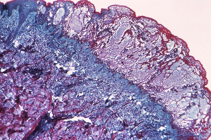 | 700 × 466 (107 KB) | Jesus Hernandez (talk | contribs) | Under a relatively low magnification of 50x, this hematoxylin-eosin (H&E)-stained photomicrograph reveals some of the cytoarchitectural histopathologic changes which you’d find in a human skin tissue specimen that included a chickenpox, or varicella ... |
You cannot overwrite this file.
File usage
The following file is a duplicate of this file (more details):
The following 2 pages use this file: