Uploads by Ssharfaei
Jump to navigation
Jump to search
This special page shows all uploaded files.
| Date | Name | Thumbnail | Size | Description | Versions |
|---|---|---|---|---|---|
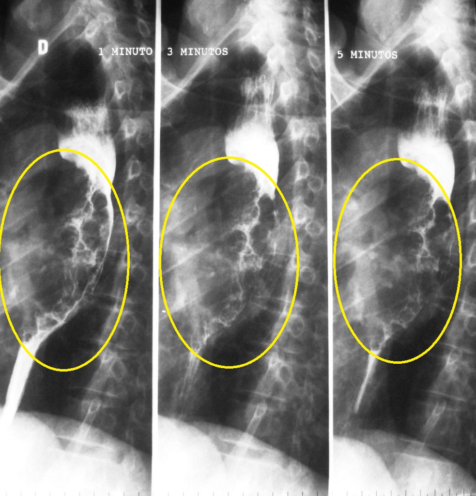
| 16:50, 13 March 2019 | Oesophageal-squamous-cell-carcinoma-2.jpg (file) |  |
243 KB | 2 | |
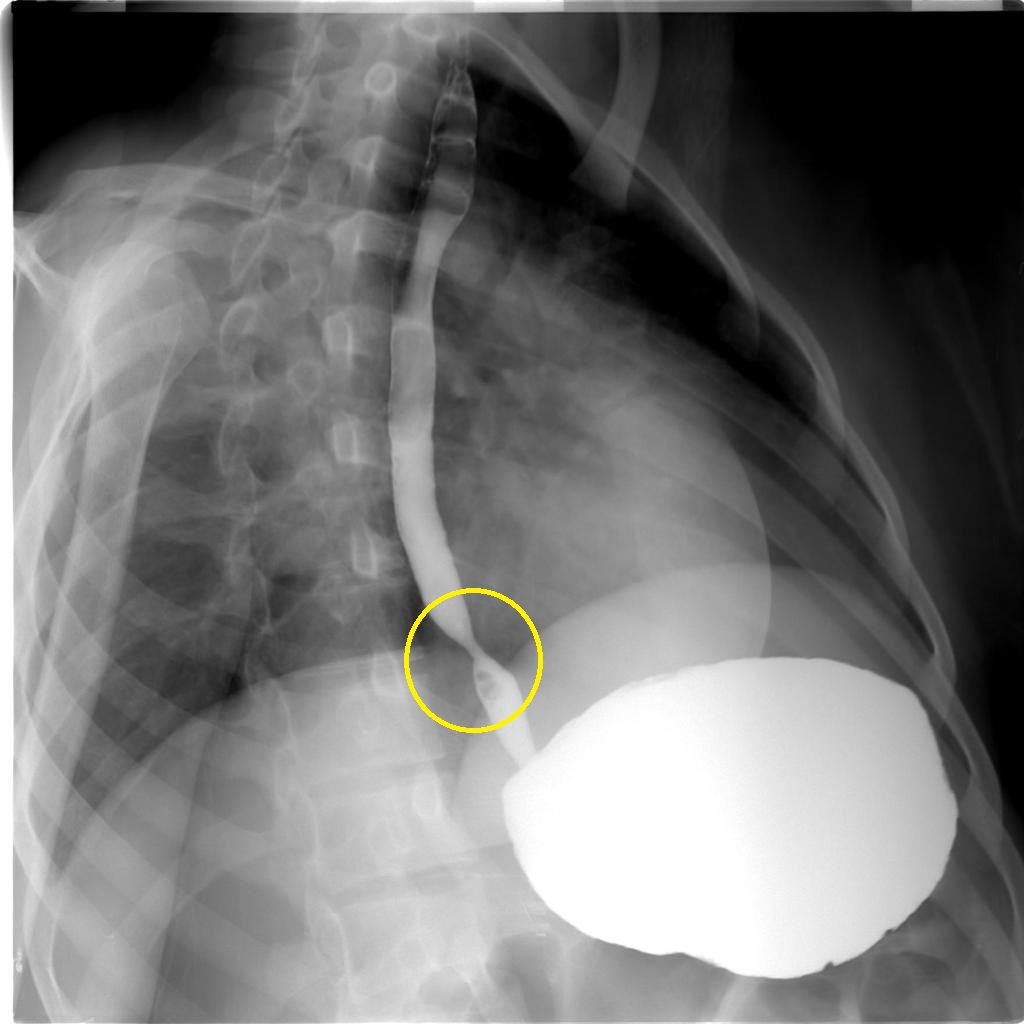
| 16:45, 13 March 2019 | Benign-oesophageal-stricture.jpg (file) |  |
102 KB | 2 | |
| 16:57, 20 June 2018 | Narrative review-Template.pdf (file) | 211 KB | 1 | ||
| 19:46, 11 May 2018 | MediaWiki Vector skin action arrow.png (file) | 231 bytes | 1 | ||
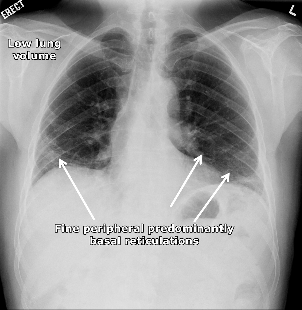
| 19:00, 3 May 2018 | 2a1be21cf39b12abeb05ff7c56aade jumbo.jpeg (file) |  |
497 KB | Patient with polymyositis and interstitial pneumonitis - Low lung volume with fine peripheral predominantly basal reticulations. Case courtesy of Dr Hani Salam, Radiopaedia.org | 1 |
| 18:54, 3 May 2018 | 34452aa9eff9ca2ec9ecdc07c4137a jumbo.jpg (file) |  |
215 KB | 1 | |
| 18:50, 3 May 2018 | 83ae133c18c2a960a5c530d2b94a0a jumbo.jpg (file) |  |
153 KB | 1 | |
| 18:49, 3 May 2018 | 96d70ca8777699049bd61c9961af78 jumbo.jpg (file) |  |
175 KB | 3 | |
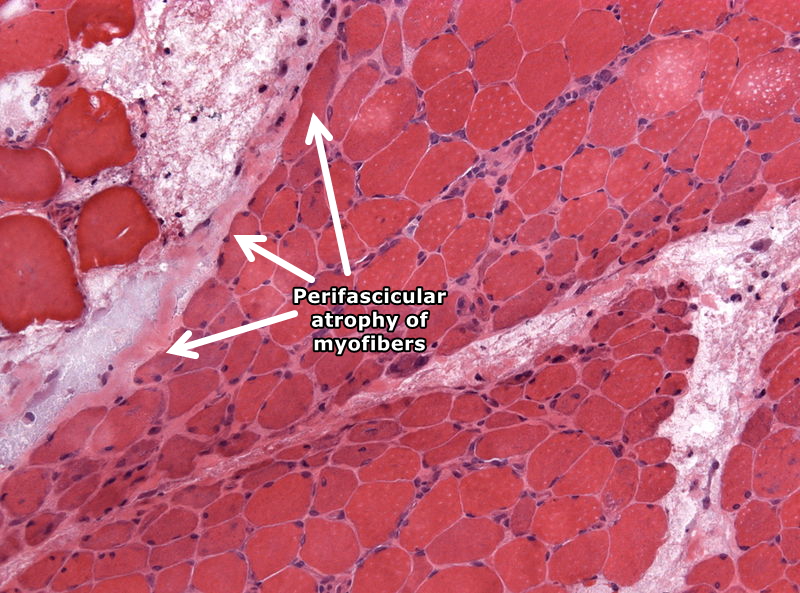
| 18:25, 3 May 2018 | 800px-NP MGMT 0252.jpg (file) |  |
282 KB | Histopathology specimen - muscle biopsy with perifascicular atrophy of myofibers as seen in dermatomyositis via librepathology.org | 1 |
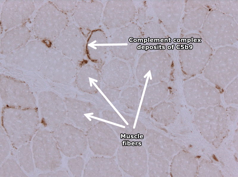
| 18:18, 3 May 2018 | 800px-Dermatomyositis c5b9.jpg (file) |  |
187 KB | Immunostain in a case of dermatomyositis displaying complement complex depositis of C5b9 Via librepathology.org | 1 |
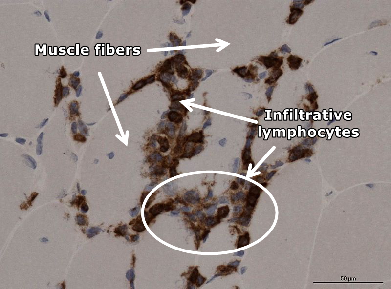
| 18:09, 3 May 2018 | 800px-Neuropathology case XII 03.jpg (file) |  |
195 KB | Muscle biopsy frozen section specimen - Myositis (CD45 stain) Partial invasion of lymphocytes within the muscle fibers via librepathology.org | 1 |
| 17:59, 3 May 2018 | Neuropathology case XII 01.jpg (file) |  |
667 KB | Muscle biopsy frozen section specimen - Myositis (HE stain)-Partial invasion of lymphocytes within the muscle fibers via librepathology.org | 1 |
| 14:41, 13 April 2018 | Polymyositis-1.JPG (file) |  |
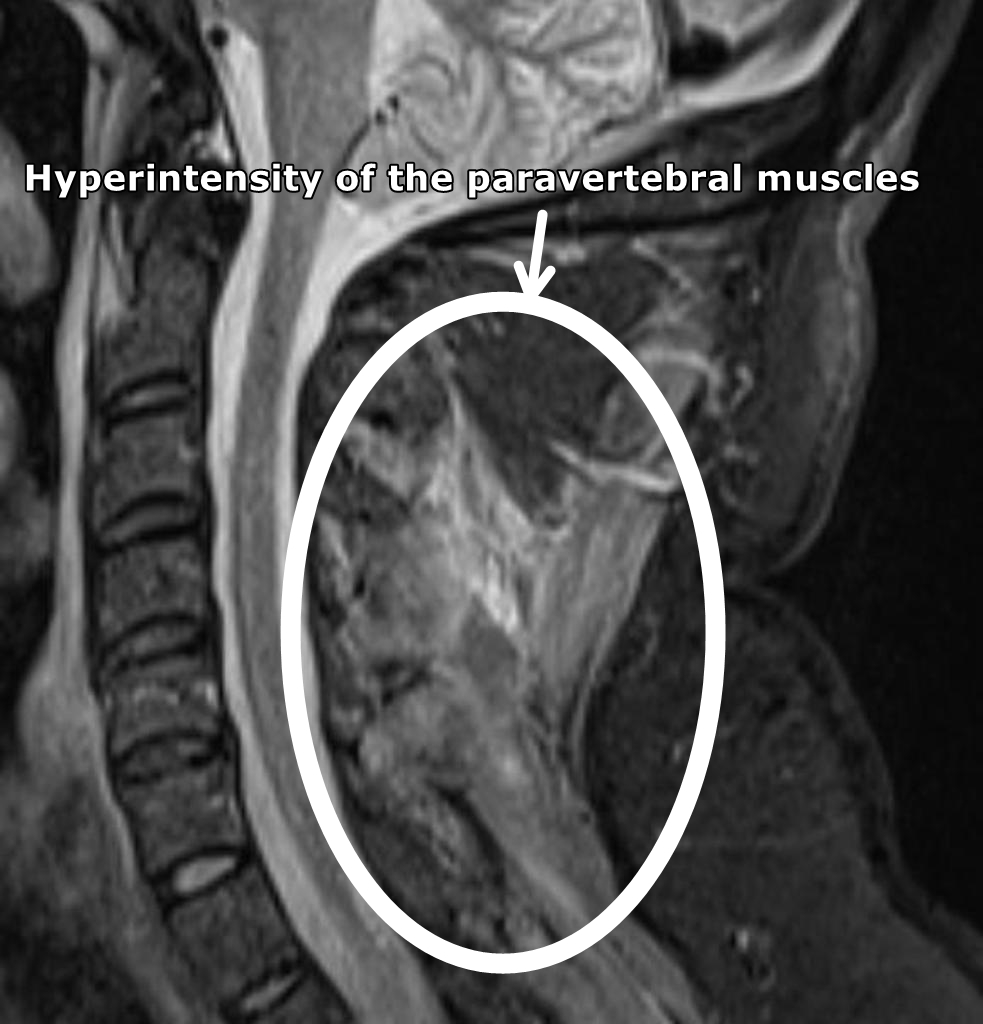
238 KB | MRI (sagittal STIR) of the cervical spine demonstrates diffuse hyperintensity of the paravertebral muscles. Case courtesy of Dr Daniela Seixas via Radiopaedia.org<ref name="urlPolymyositis | Radiology Case | Radiopaedia.org">{{cite web |url=https://ra... | 1 |
| 19:33, 3 April 2018 | Dermatomyositis5.jpg (file) |  |
399 KB | Dermatomyositis, Gottron's papules. Erythematous plaques overlying the elbows in two patients with juvenile dermatomyositis. In some patients, small erythematous plaques may overly the extensor aspects of larger joints, such as the elbows, knees, and m... | 1 |
| 01:55, 31 March 2018 | Aspiration pneumonia201711-3264.jpg (file) |  |
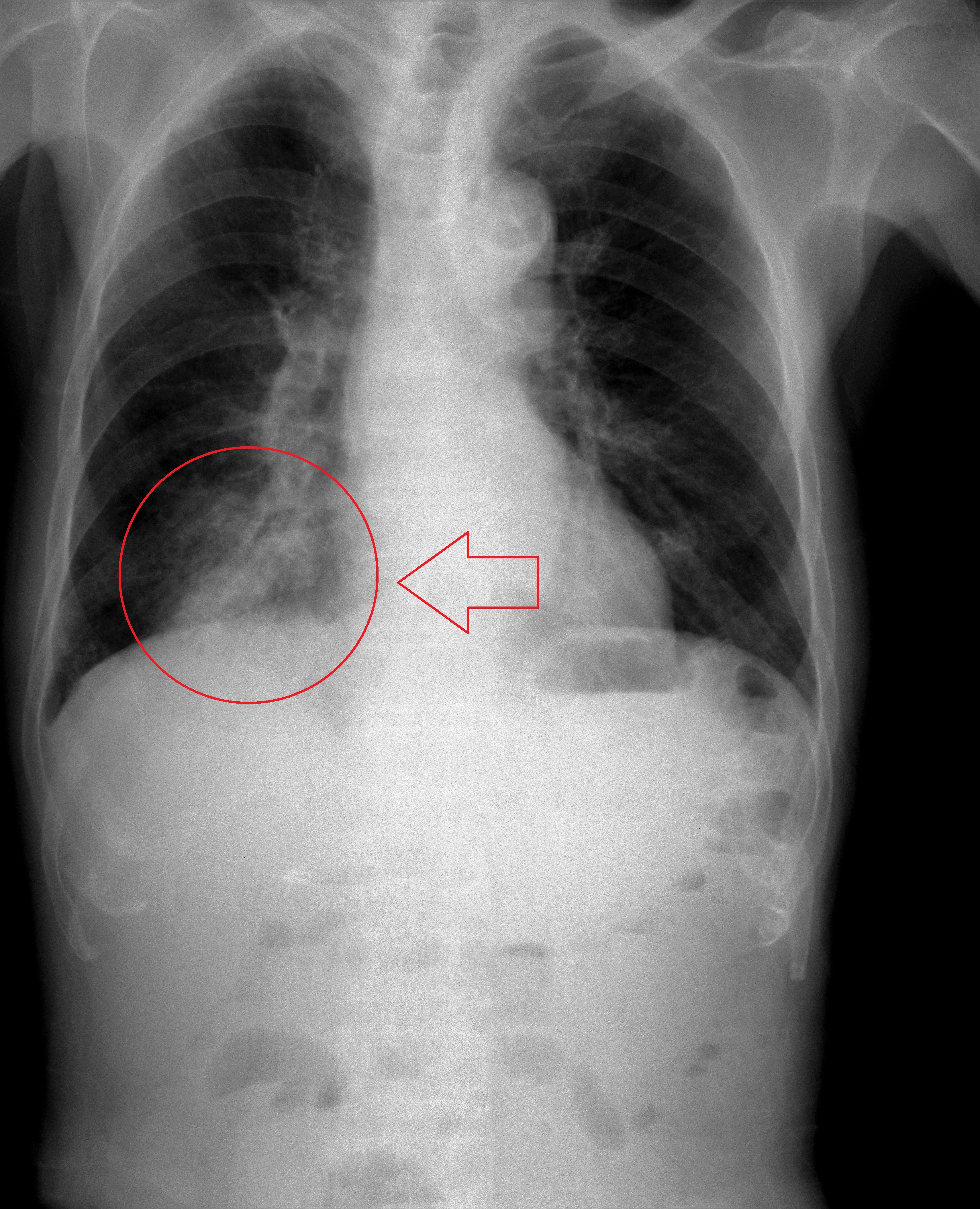
4.51 MB | Aspiration pneumonia. 83y.o. male. Right side lower lobe. By melvil - Own work, CC BY-SA 4.0, via wikimedia<ref name="urlFile:Aspiration pneumonia201711-3264.jpg - Wikimedia Commons">{{cite web |url=+https://commons.wikimedia.org/w/index.php?curid=6465... | 1 |
| 01:50, 31 March 2018 | Lipid pneumonia, exogenous (3791887936).jpg (file) | .jpg) |
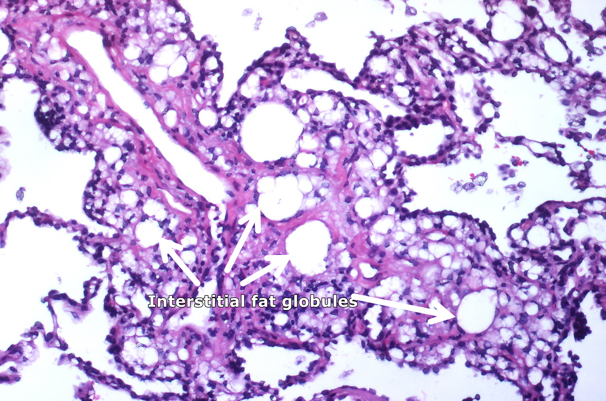
1.81 MB | Numerous interstitial fat globules of varying size accompanied by inflammation and fibrosis is characterstic of chronic lipid pneumonia secondary to lipid aspiration. By Yale Rosen from USA - Lipid pneumonia, exogenousUploaded by CFCF, CC BY-SA 2.0, Vi... | 1 |
| 01:46, 31 March 2018 | Kayexalate aspiration Case 125 (4692318776).jpg (file) | .jpg) |
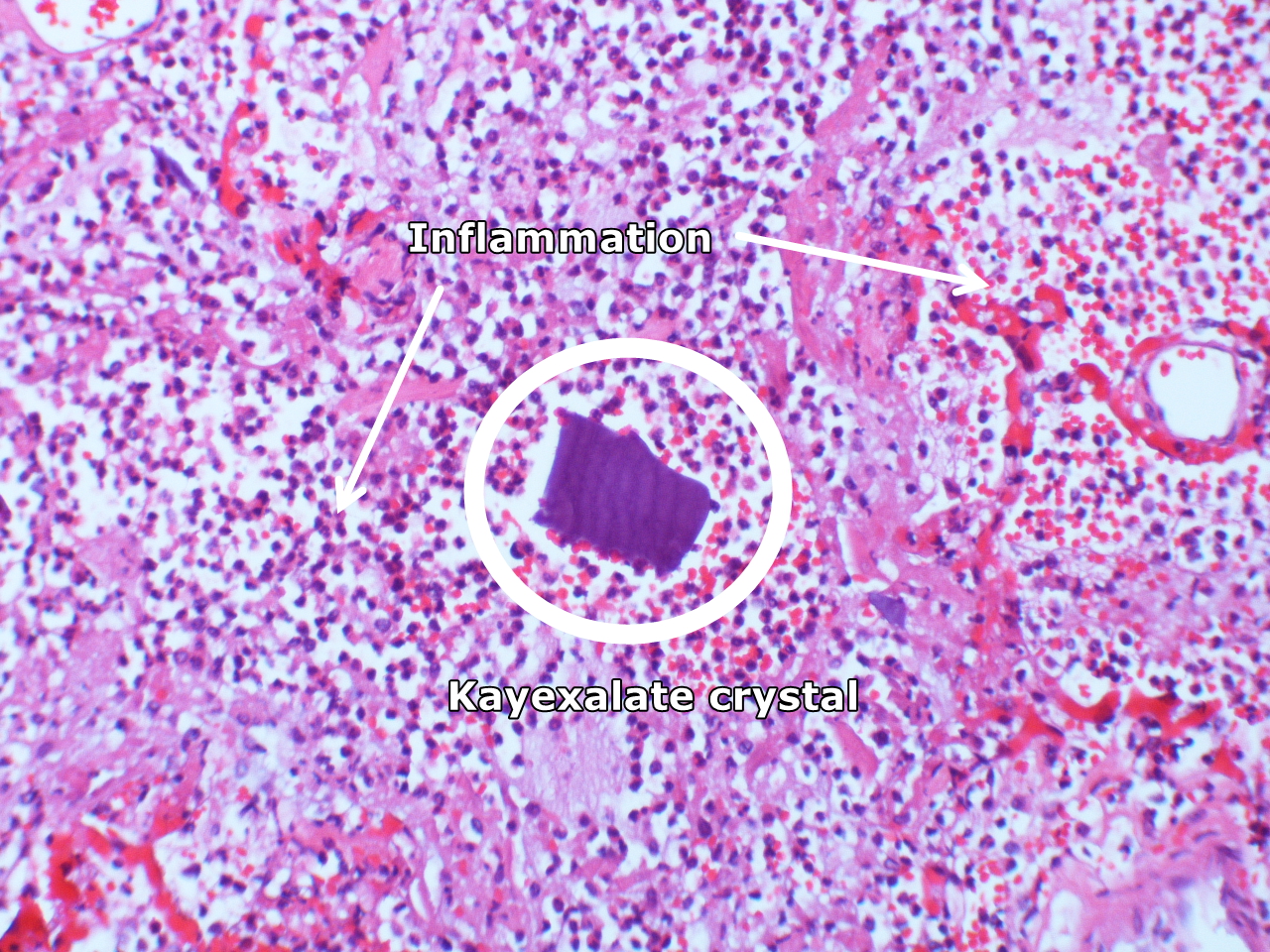
1.35 MB | Intraalveolar kayexalate crystal; acute pneumonitis. By Yale Rosen from USA - Kayexalate aspiration Case 125Uploaded by CFCF, CC BY-SA 2.0, Via Wikimedia<ref name="urlFile:Kayexalate aspiration Case 125 (4692318776).jpg - Wikimedia Commons">{{cite web... | 1 |
| 01:41, 31 March 2018 | Aspiration pneumonia (5613146123).jpg (file) | .jpg) |
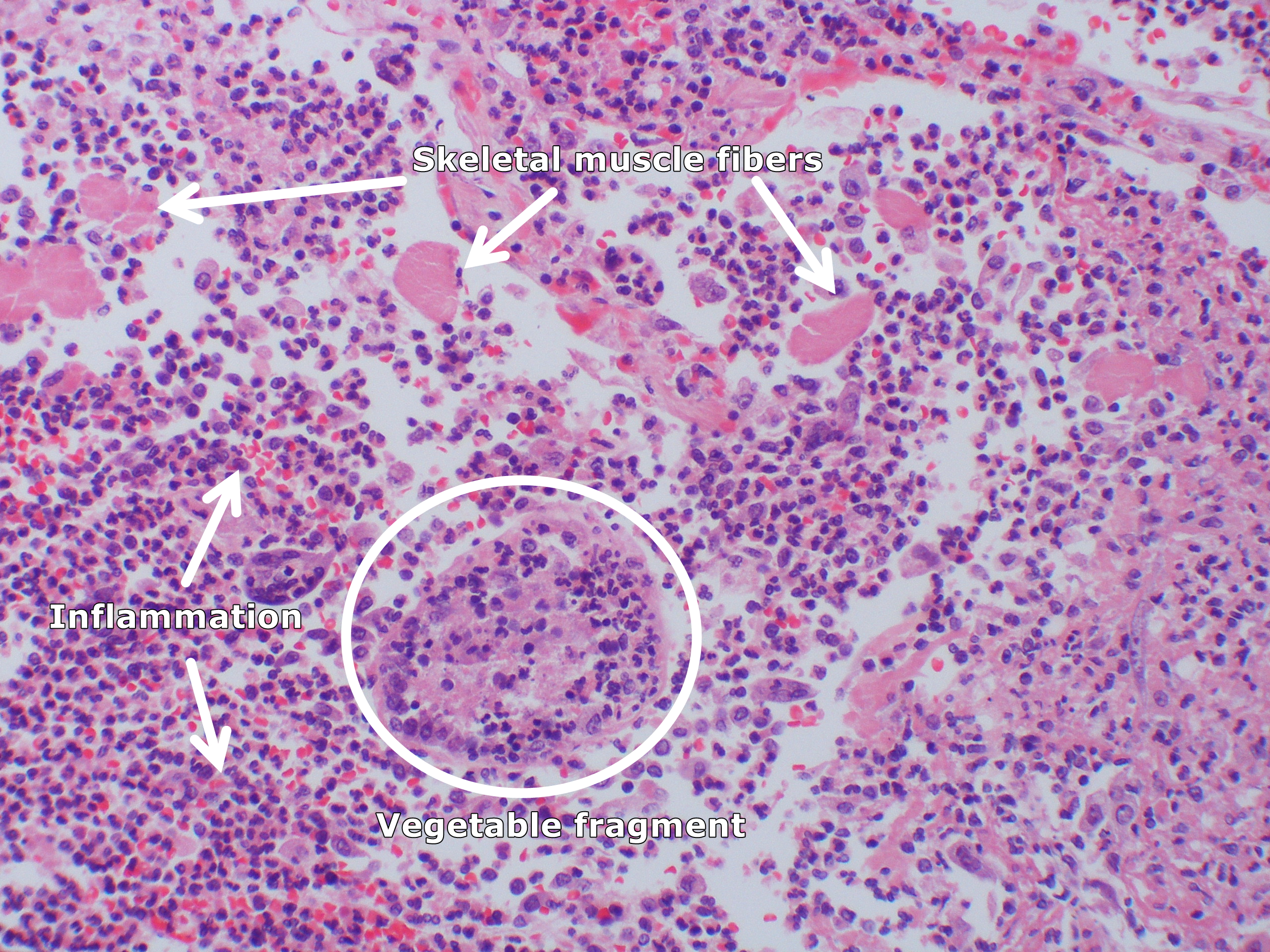
3 MB | Acute aspiration pneumonia with numemous skeletal muscle fibers and a vegetable fragment infiltrated by polys. By Yale Rosen from USA - Aspiration pneumoniaUploaded by CFCF, CC BY-SA 2.0, Via Wikimedia<ref name="urlFile:Aspiration pneumonia (5613146123... | 1 |
| 01:28, 31 March 2018 | Aspiration pneumonia (5613726286).jpg (file) | .jpg) |
2.84 MB | Aspirated vegetable material surrounded by macrophages. This structure has a thick outer wall composed of cellulose surrounding a latticework of individual cells with thick cell walls composed of cellulose. By Yale Rosen from USA - Aspiration pneumonia... | 1 |
| 01:10, 31 March 2018 | Aspiration (4858360012).jpg (file) | .jpg) |
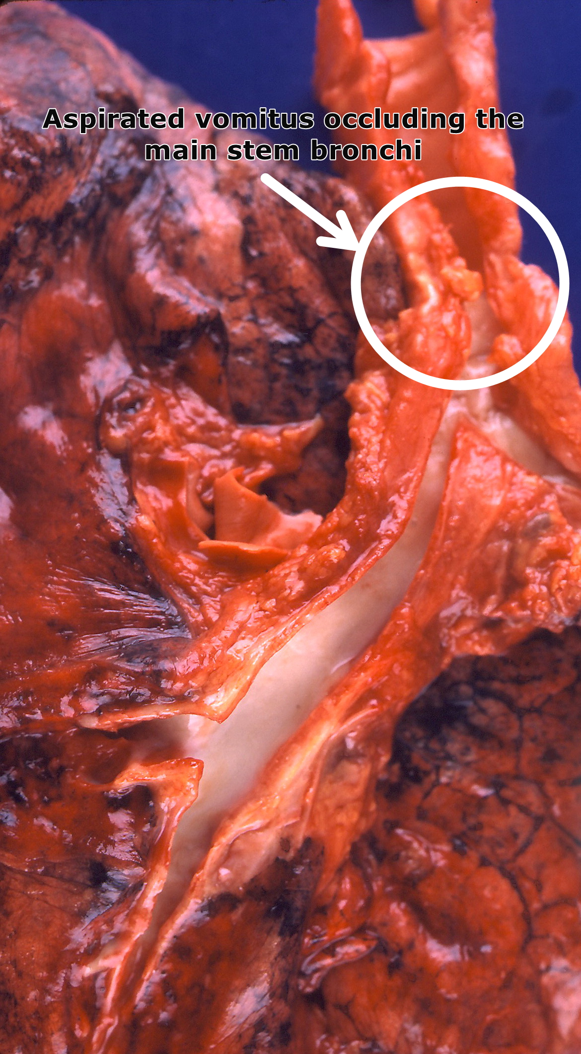
1.28 MB | Aspirated vomitus occluding the main stem bronchi. By Yale Rosen from USA - AspirationUploaded by CFCF, CC BY-SA 2.0, Via Wikimedia<ref name="urlFile:Aspiration (4858360012).jpg - Wikimedia Commons">{{cite web |url=https://commons.wikimedia.org/w/index... | 1 |
| 00:20, 31 March 2018 | Aspirated corn kernel (3791886968).jpg (file) | .jpg) |
1.97 MB | Aspirated corn kernel By Yale Rosen from USA - Uploaded by CFCF, CC BY-SA 2.0, Via Wikimedia<ref name="urlFile:Aspirated corn kernel (3791886968).jpg - Wikimedia Commons">{{cite web |url=https://commons.wikimedia.org/w/index.php?curid=31128322 |title=F... | 1 |
| 21:43, 12 February 2018 | 2119 Pulmonary Circuit.jpg (file) |  |
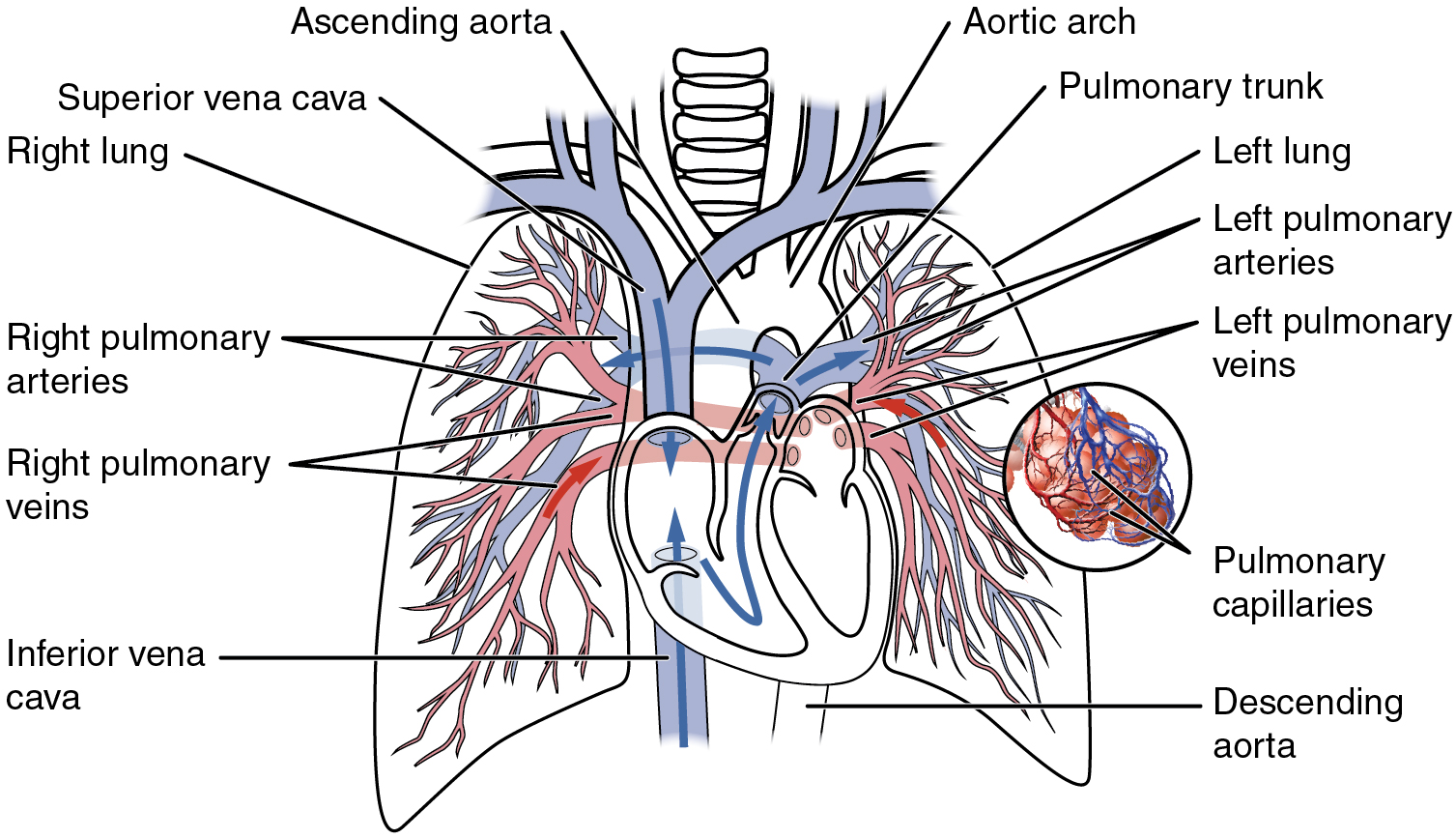
665 KB | Illustration from Anatomy & Physiology, Connexions Web site. http://cnx.org/content/col11496/1.6/, Jun 19, 2013. By OpenStax College - Anatomy & Physiology, Connexions Web site. http://cnx.org/content/col11496/1.6/, Jun 19, 2013., CC BY 3.0,<ref name="... | 1 |
| 21:40, 12 February 2018 | Pulmonary Blood Circulation.png (file) |  |
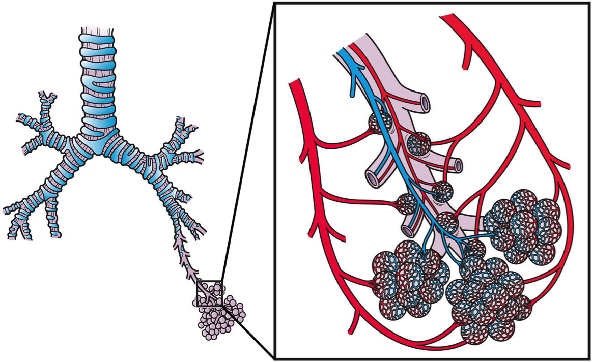
776 KB | This slide shows the arterial and venous blood circulation of the pulmonary system. By Artwork by Holly Fischer - http://open.umich.edu/education/med/resources/second-look-series/materials - Respiratory Tract Slide 20, CC BY 3.0,<ref name="urlFile:Pulm... | 1 |
| 16:48, 31 January 2018 | Coronal CT - FAP.jpg (file) |  |
111 KB | Coronal CT scan shows multiple filling defects in a patient with familial adenomatous polyposis. Case courtesy of Dr David Cuete, Radiopaedia.org | 1 |
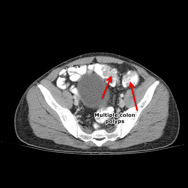
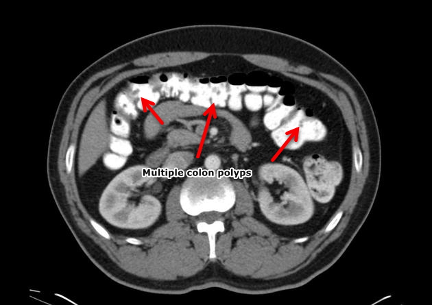
| 16:47, 31 January 2018 | Axial CT- FAP.jpg (file) |  |
90 KB | Axial CT scan shows multiple filling defects in a patient with familial adenomatous polyposis. Case courtesy of Dr David Cuete, Radiopaedia.org | 1 |
| 16:42, 31 January 2018 | Sagittal CT - FAP.jpg (file) |  |
73 KB | Sagittal CT shows multiple filling defects in a patient with familial adenomatous polyposis. Case courtesy of Dr David Cuete, Radiopaedia.org | 1 |
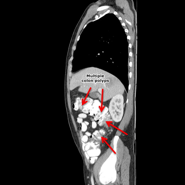
| 15:03, 31 January 2018 | Sagittal CT Colon polyps.jpeg (file) |  |
49 KB | Sagittal CT scan shows colon polyp | 1 |
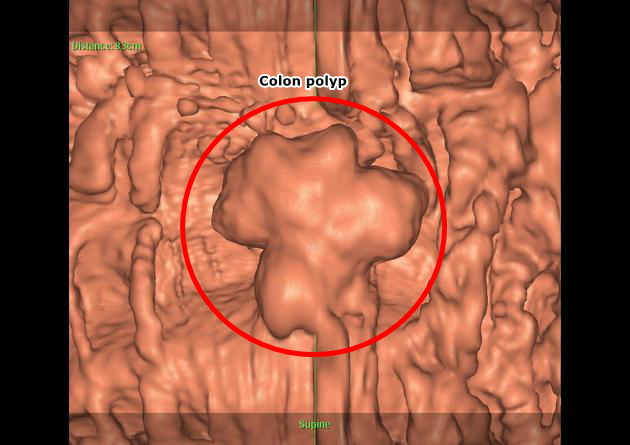
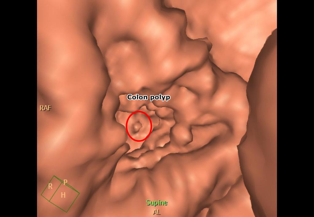
| 15:01, 31 January 2018 | Virtual colonoscopy Colon polyp.jpeg (file) |  |
116 KB | Virtual colonoscopy shows pedunculated colon polyp-Case courtesy of Dr Ayaz Hidayatov, Radiopaedia.org | 1 |
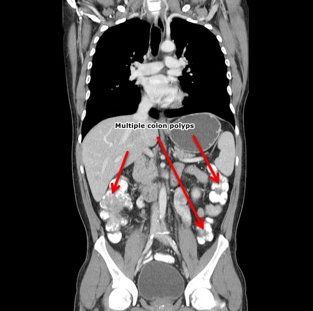
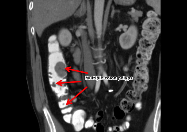
| 14:56, 31 January 2018 | Coronal CT colon polyps.jpeg (file) |  |
69 KB | Coronal CT scan shows multiple pedunculated colon polyps-Case courtesy of Dr Ayaz Hidayatov, Radiopaedia.org | 1 |
| 14:51, 31 January 2018 | 6fdbe648a73ff963226cfcf5c0414a big gallery.jpeg (file) |  |
76 KB | 1 | |
| 14:22, 31 January 2018 | Colonic-polyp-virtual-colonoscopy-1 (0).jpg (file) | .jpg) |
156 KB | 1 | |
| 15:59, 30 January 2018 | Familial adenomatous polyposis.jpg (file) |  |
78 KB | 1 | |
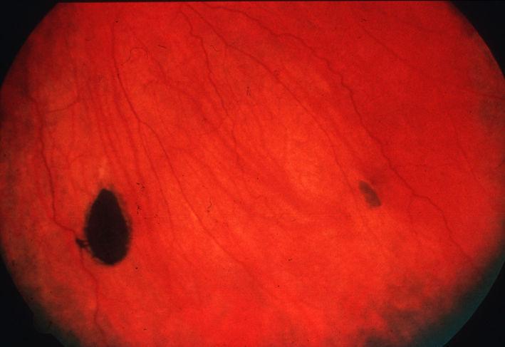
| 15:50, 30 January 2018 | CHRPE Congenital hypertrophy of the retinal pigment epithelium.jpg (file) |  |
31 KB | 1 | |
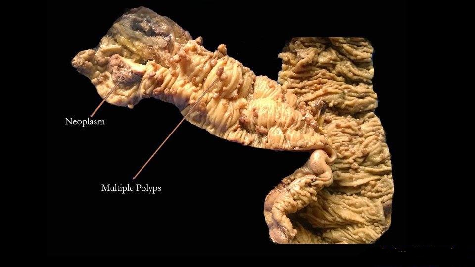
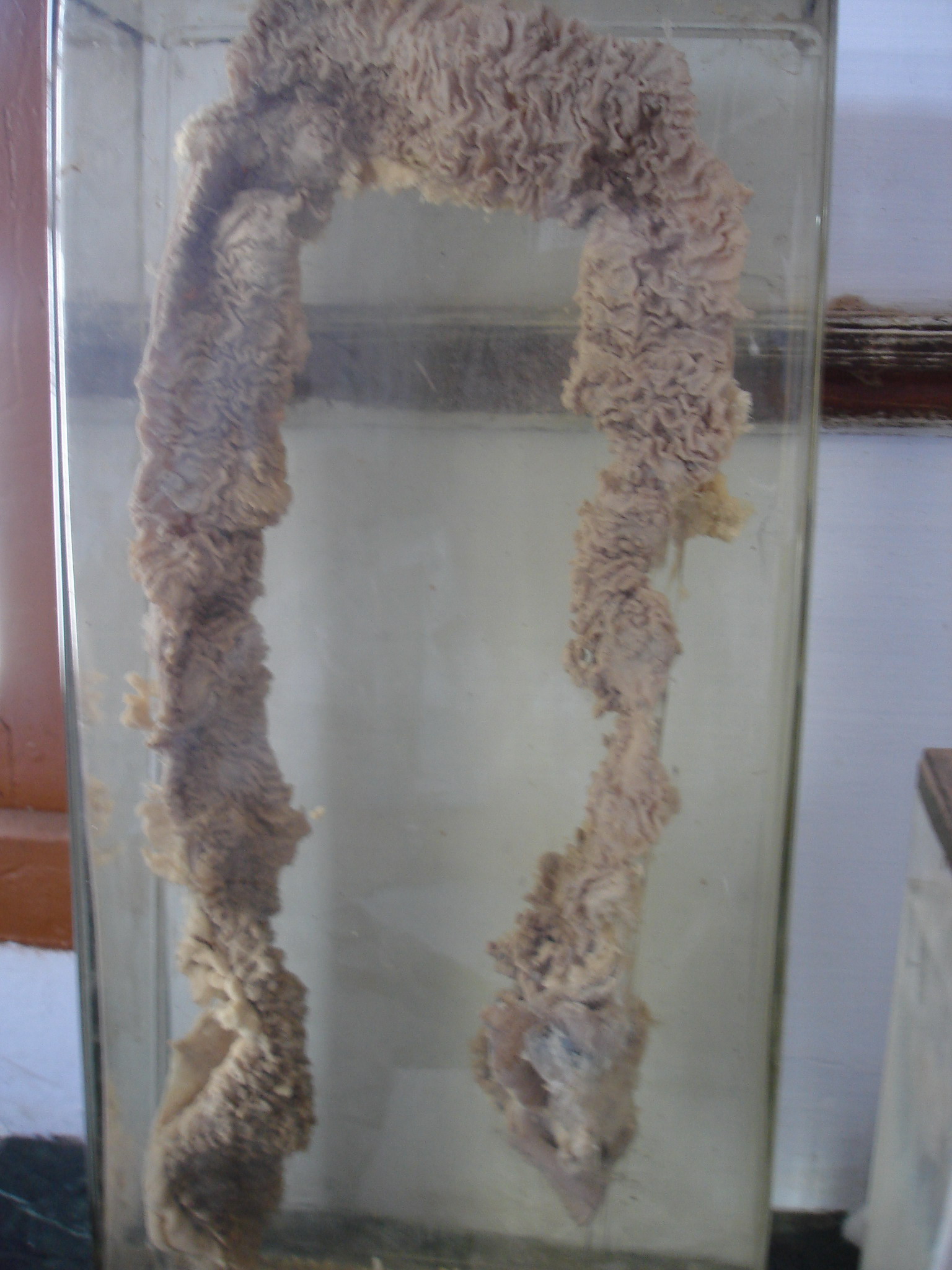
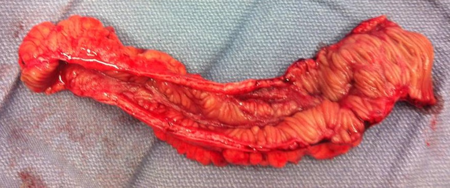
| 15:30, 30 January 2018 | Familial Adenomatous Polyposis intestine.jpg (file) |  |
497 KB | 2 | |
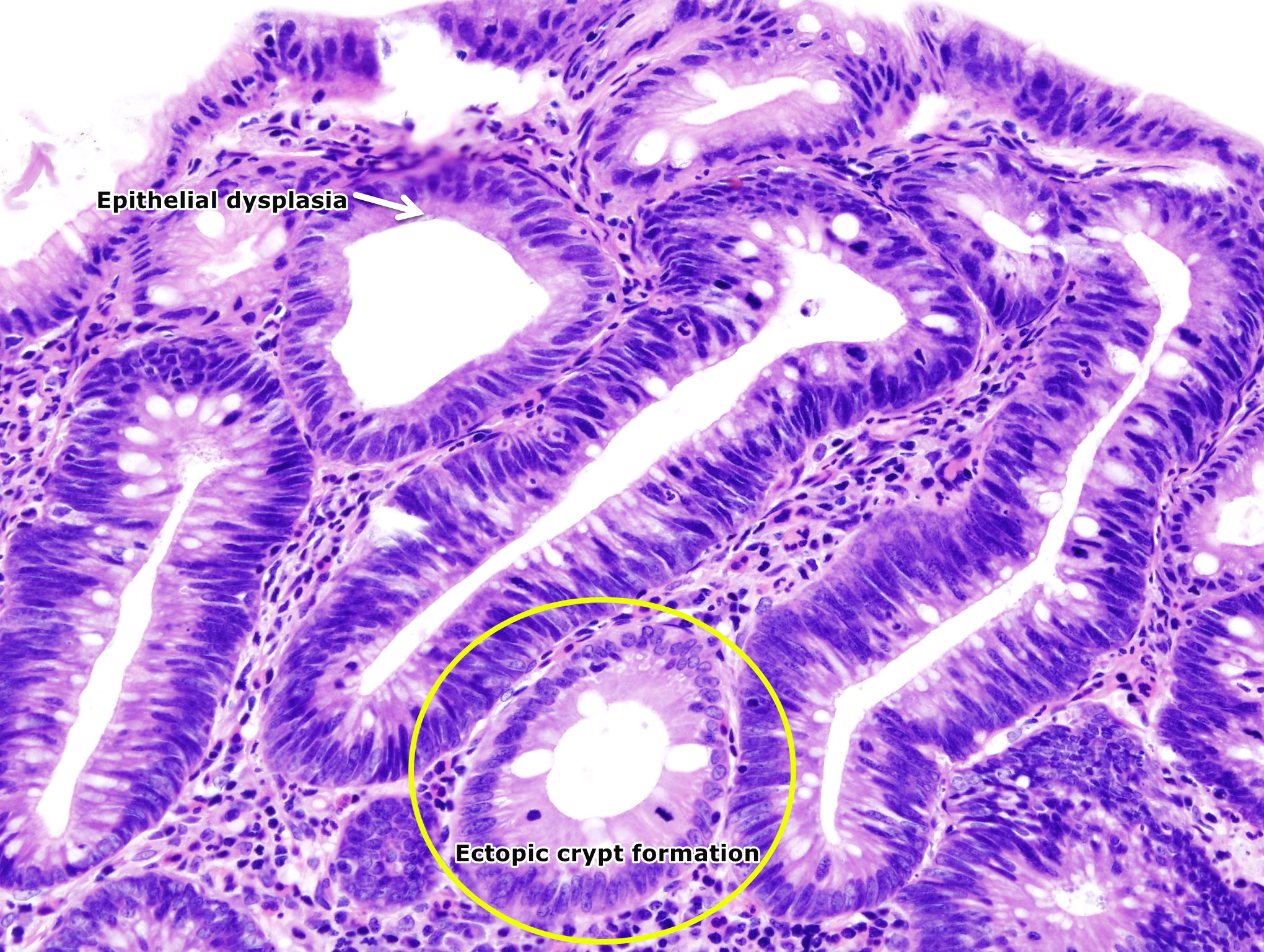
| 21:14, 26 January 2018 | Colon adenoma (1).jpg (file) | .jpg) |
1.81 MB | 3 | |
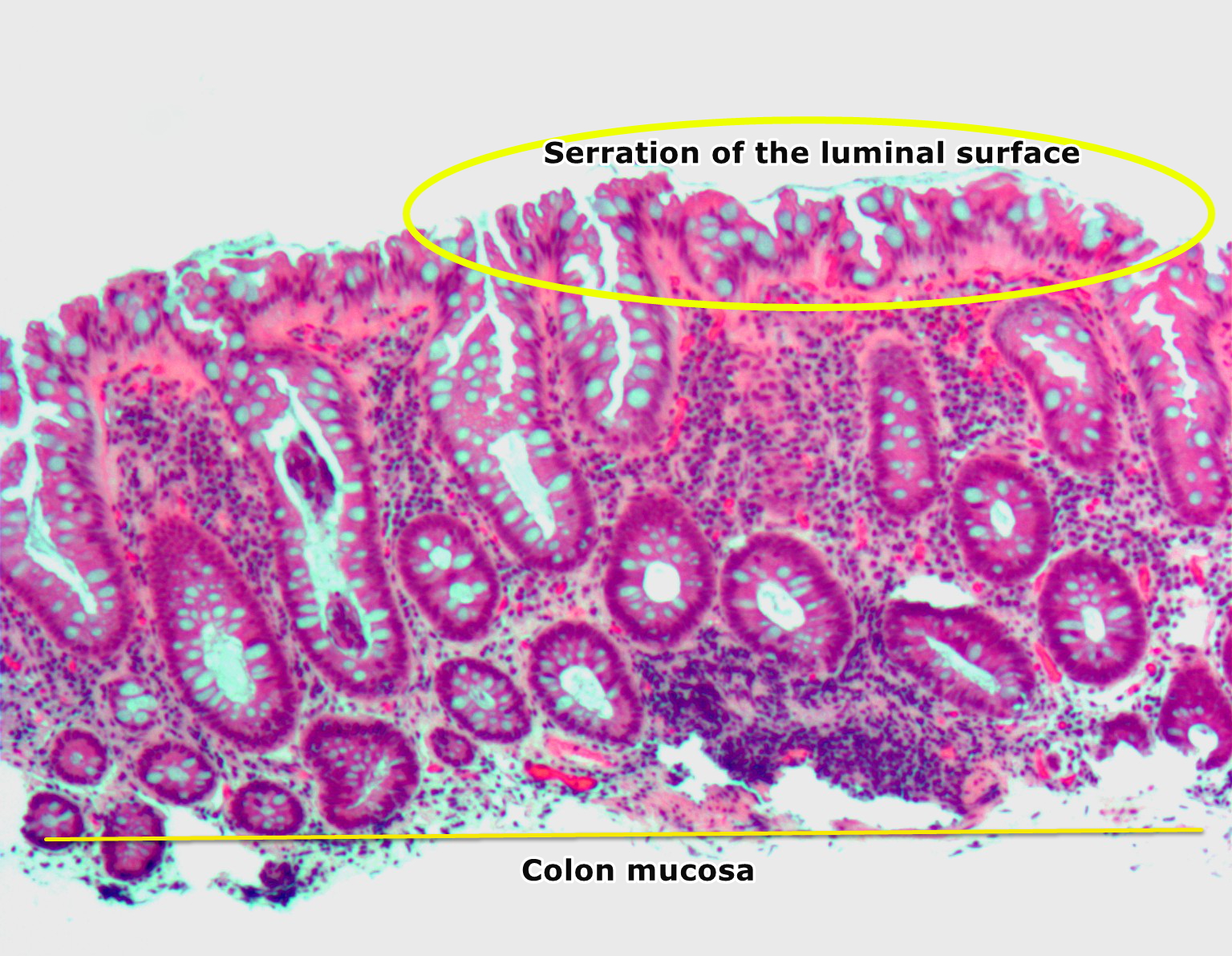
| 21:02, 26 January 2018 | Hyperplastic polyp2.jpg (file) |  |
946 KB | 3 | |
| 17:58, 23 January 2018 | Villous adenoma of the sigmoid colon, gross pathology.jpg (file) |  |
64 KB | 1 | |
| 17:43, 23 January 2018 | Colon-Polyp.jpg (file) |  |
778 KB | 1 | |
| 17:39, 23 January 2018 | Hyperplastic polyp of the colon, HE.png (file) |  |
1.93 MB | 1 | |
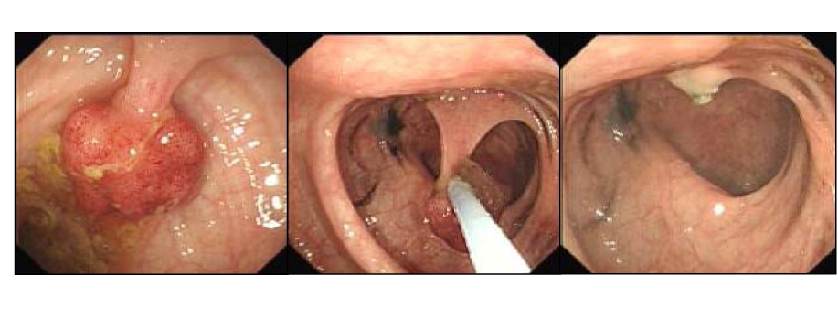
| 17:12, 23 January 2018 | Polypectomy.jpg (file) |  |
185 KB | 1 | |
| 17:12, 20 December 2017 | Image of resected colon segment with cancer & 4 nearby polyps plus schematic of field defects with sub-clones.jpg (file) |  |
625 KB | 1 | |
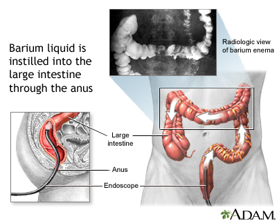
| 20:55, 19 December 2017 | Barium enema.jpg (file) |  |
81 KB | 1 | |
| 16:09, 5 December 2017 | Diagram of the small bowel 01 CRUK 045.jpg (file) |  |
254 KB | Diagram of the small bowel 01. Source: Wikimedia.org By Cancer Research UK - Original email from CRUK, CC BY-SA 4.0, https://commons.wikimedia.org/w/index.php?curid=34332940 | 1 |
| 14:32, 5 December 2017 | Crohn Jejunum.png (file) |  |
300 KB | Partial jejunum affected by morbus Crohn. Source: Wikimedia.org By Jaroslav Cehovsky - Camera, Public Domain, https://commons.wikimedia.org/w/index.php?curid=1458390 | 1 |
| 15:19, 4 December 2017 | ResectedIleum.jpg (file) |  |
137 KB | Terminal ileum resected for Crohn's disease. By PPSE15 - Own work, CC BY-SA 4.0<ref name="urlFile:ResectedIleum.jpg - Wikimedia Commons">{{cite web |url=https://commons.wikimedia.org/w/index.php?curid=39360128 |title=File:ResectedIleum.jpg - Wikimedia... | 1 |
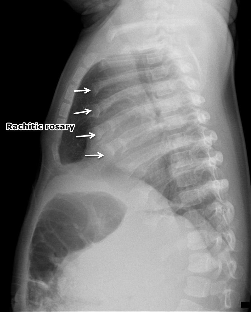
| 01:04, 26 November 2017 | Rachitic-rosary (1).png (file) | .png) |
433 KB | Rachitic rosary: Widening of the anterior rib ends at the costochondral junctions. Case courtesy of Dr Dalia Ibrahim, Radiopaedia.org, rID: 47584 https://radiopaedia.org/cases/47584 | 1 |
| 01:04, 26 November 2017 | Rachitic-rosary.png (file) |  |
529 KB | Rachitic rosary: Widening of the anterior rib ends at the costochondral junctions. Case courtesy of Dr Dalia Ibrahim, Radiopaedia.org, rID: 47584 https://radiopaedia.org/cases/47584 | 1 |
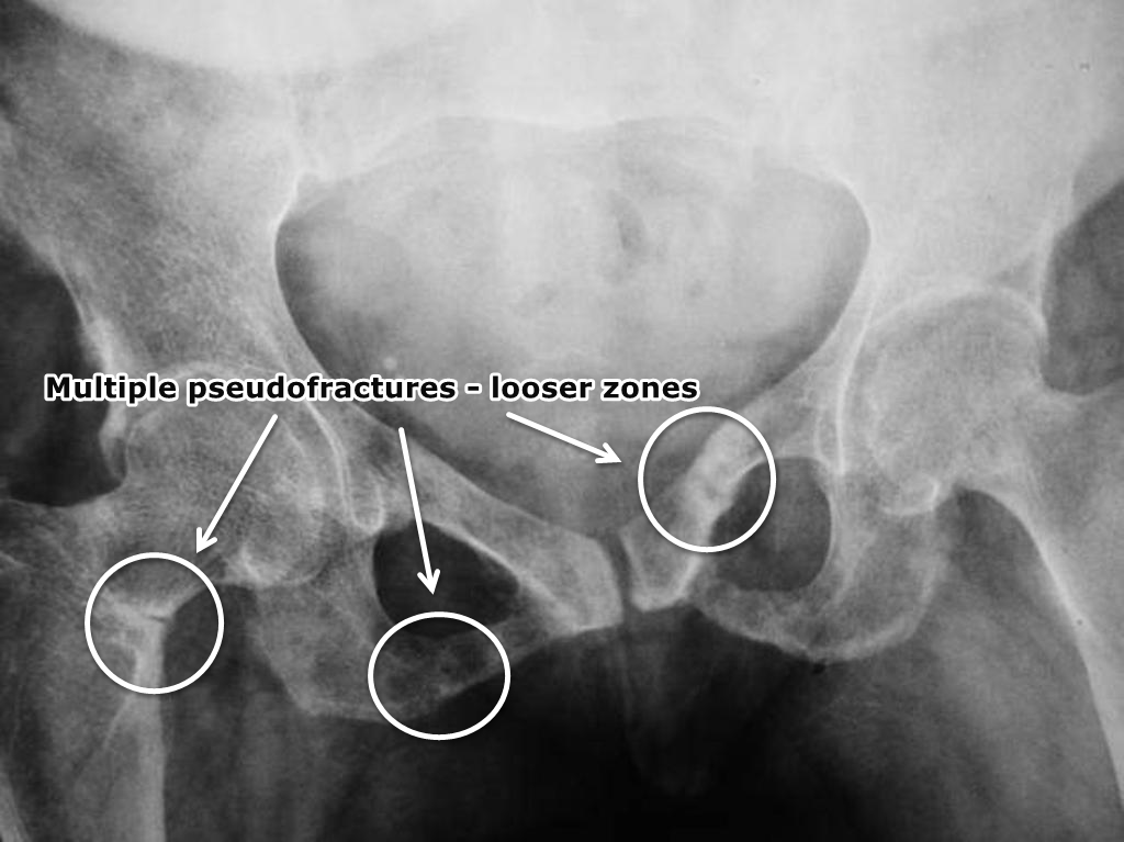
| 00:53, 26 November 2017 | Osteomalacia-looser-zones-1.png (file) |  |
452 KB | Osteopenic changes involving bony pelvis and proximal femurs. Multiple pseudofractures/Looser zones are seen involving superior and inferior pubic rami bilaterally. There is also a transcervical fracture on the right side. Case courtesy of Dr Iqbal Na... | 1 |
| 23:57, 25 November 2017 | Rickets-2.png (file) |  |
829 KB | Case courtesy of A.Prof Frank Gaillard, Radiopaedia.org, rID: 8225 https://radiopaedia.org/cases/8225 The physes are widened with metaphyseal flaring. Note how the bones are coarse. | 1 |
| 23:51, 25 November 2017 | Rickets-with-pathological-fracture.png (file) |  |
204 KB | Pathological fracture of the femur shaft. The fracture seems weeks old with beginning callus. however, it is only partially consolidated. The main impression is the saber-like appearance of the bone which is common for patients with vitamin D deficienc... | 1 |