Eardrum
Editor-In-Chief: C. Michael Gibson, M.S., M.D. [1]
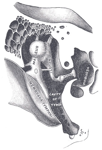
The tympanic membrane, colloquially known as the eardrum, is a thin membrane that separates the external ear from the middle ear. Its function is to transmit sound vibrations from the air, conducted through the external acoustic meatus to the ossicles inside the middle ear. The malleus bone bridges the gap between the eardrum and the other ossicles.
Arterial supply - outer surface is supplied by the deep auricular branch of the maxillary artery,inner surface is supplied by the anterior tympanic branch of the maxillary artery & by the posterior tympanic branch of the stylomastoid branch of the posterior auricular artery. Venous drainage - outer surface drains into the external jugular vein.inner surface drains into the transverse sinus & into the venous plexus around the auditory tube.
Lymphatic drainage – pass to the preauricular & retropharyngeal nodes. Nerve supply – Outer surface – anteroinferior part is supplied by the auriculotemporal nerve & the posterosuperior part by the auricular branch of the vagus nerve. Inner surface is supplied by the tympanic branch of the glossopharyngeal nerve through the tympanic plexus.
Rupture or perforation of the eardrum can lead to conductive hearing loss.
Development
The eardrum forms from the joining of the expanding first pharyngeal pouch and groove. Around day 30 of gestation, the endoderm-lined first expands to form the tympanic cavity, which subsequently envelops the inner ear ossicles. Simultaneously, the first pharyngeal groove, which is lined with ectoderm, expands to form the developing external auditory meatus. Separated by a thin layer of splanchnic mesoderm, the tympanic cavity and external auditory meatus join to form the tympanic membrane. As a result, the tympanic membrane is one of very few adult structures that is derived from all three germ layers. The skin that covers the outer surface of the tympanic membrane is derived from ectoderm, the fibrous tissue that forms the actual membrane is derived from mesoderm, and the mucus membrane that lines the inner surface of the membrane is derived from endoderm.
Clinical Aspects
When examining the tympanic membrane with an otoscope, a bright cone of light is seen in the anterior-inferior part of the membrane. This light is known as the "cone of light" or "light reflex". The tympanic membrane is separated into four quadrants, with the center of the four quadrants being the umbo. Nerves, specifically the chorda tympani nerve, and arteries pass through the layers of the superior portion of the membrane. Thus, when the tympanic membrane needs to be incised for medical procedures, ENT surgeons will always cut through the inferior and posterior part of the membrane to avoid the vasculature, nerves, and bones associated with the membrane.
Additional images
-
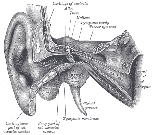 External and middle ear, opened from the front. Right side.
 -
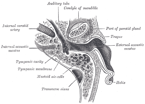 Horizontal section through left ear; upper half of section.
 -
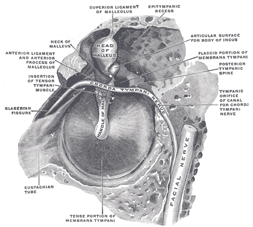 The right membrana tympani with the hammer and the chorda tympani, viewed from within, from behind, and from above.
 -
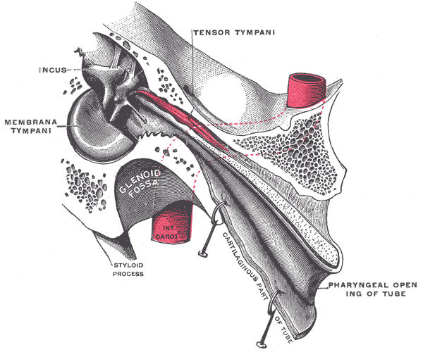 Auditory tube, laid open by a cut in its long axis.
 -
 Chain of ossicles and their ligaments, seen from the front in a vertical, transverse section of the tympanum.


External links
de:Trommelfell et:Trummikile id:Gendang telinga it:Timpano (anatomia) he:עור התוף la:Membrana tympani lt:Būgnelis nl:Trommelvlies no:Trommehinne nn:Trommehinne sk:Bubienok (cicavce) sr:Бубна опна fi:Tärykalvo uk:Барабанна перетинка