File:Ossifying fibroma.jpg
Ossifying_fibroma.jpg (437 × 270 pixels, file size: 20 KB, MIME type: image/jpeg)
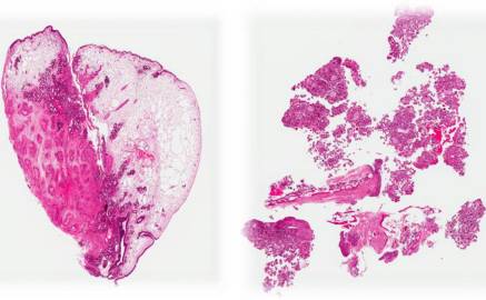
Summary
Histopathologicaly, this lesion is characterized by spindle cell proliferation packed with globular cementum droplets and round/ovoid fragments of woven bone. These spindle cells appear benign and associated with psammomatoid cementum droplets and some woven bone and consistent with aggressive ossifying fibroma, psammomatous type IV,Alghonaim Y, ALRashed ALHumaid S, Arafat A. Aggressive ossifying fibroma of right ethmoidal sinus: A case report. Int J Surg Case Rep. ;53:513–516. doi:10.1016/j.ijscr.2017.12.026,https://www.ncbi.nlm.nih.gov/pmc/articles/PMC6290393/
File history
Click on a date/time to view the file as it appeared at that time.
| Date/Time | Thumbnail | Dimensions | User | Comment | |
|---|---|---|---|---|---|
| current | 19:24, 29 May 2019 | | 437 × 270 (20 KB) | Maneesha Nandimandalam (talk | contribs) | Histopathologicaly, this lesion is characterized by spindle cell proliferation packed with globular cementum droplets and round/ovoid fragments of woven bone. These spindle cells appear benign and associated with psammomatoid cementum droplets and some... |
You cannot overwrite this file.
File usage
The following page uses this file: