File:MRI pilocytic astrocytoma 3.jpg

MRI_pilocytic_astrocytoma_3.jpg (547 × 181 pixels, file size: 39 KB, MIME type: image/jpeg)
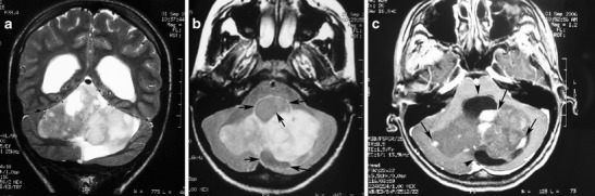
A pilocytic astrocytoma with a predominantly solid mass and a minimal cyst-like component in a 14-year-old girl. a This coronal T2-weighted image shows a large, well-marginated mass, involving the vermis and both cerebellar hemispheres that effaces the fourth ventricle. b On an axial FLAIR image, the solid mass shows high signal intensity with hypointense cystic areas (arrows). c On an axial contrast-enhanced image, the mass shows inhomogeneous contrast enhancement, with nodules that exhibit intense enhancement (arrows) and other cystic areas that remain unenhanced (arrowheads).
File history
Click on a date/time to view the file as it appeared at that time.
| Date/Time | Thumbnail | Dimensions | User | Comment | |
|---|---|---|---|---|---|
| current | 17:42, 29 October 2015 | 547 × 181 (39 KB) | Sujit Routray (talk | contribs) | A pilocytic astrocytoma with a predominantly solid mass and a minimal cyst-like component in a 14-year-old girl. '''a''' This coronal T2-weighted image shows a large, well-marginated mass, involving the vermis and both cerebellar hemispheres that effac... | |
| 17:42, 29 October 2015 | 547 × 181 (39 KB) | Sujit Routray (talk | contribs) | A pilocytic astrocytoma with a predominantly solid mass and a minimal cyst-like component in a 14-year-old girl. '''a''' This coronal T2-weighted image shows a large, well-marginated mass, involving the vermis and both cerebellar hemispheres that effac... |
You cannot overwrite this file.
File usage
There are no pages that use this file.