File:Legionella microscopy 2.jpg
Legionella_microscopy_2.jpg (700 × 475 pixels, file size: 82 KB, MIME type: image/jpeg)
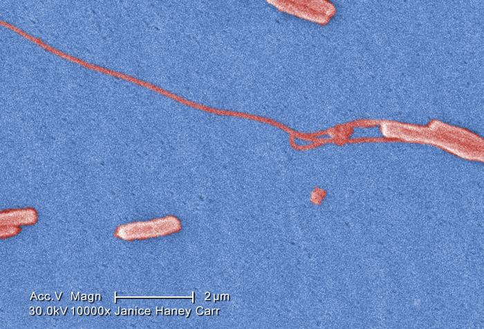
Under a very very high magnification of 10000X, this colorized scanning electron micrograph (SEM) depicted a number of Gram-negative Legionella pneumophila bacteria. Please see PHIL 11092 through 11152 for additional SEMs of these organisms, specifically PHIL 11145 for a black and white version of this image. Of particular importance, is the presence of polar flagella, and pili, or long streamers, which due to their fragile nature, in some of these views seem to be dissociated from any of the bacteria. You’ll note that a number of these bacteria seem to display an elongated-rod morphology. L. pneumophila are known to most frequently exhibit this configuration when grown in broth, however, they can also elongate when plate-grown cells age, as it was in this case, especially when they’ve been refrigerated. The usual L. pneumophila morphology consists of stout, “fat” bacilli, which is the case for the vast majority of the organisms depicted here. These bacteria originated on a 1 week-old culture plate (+/- 1 day), which had incubated a single colony, at 37oC upon a buffered charcoal yeast extract (BCYE) medium with no antibiotics.
Photo Credit: Janice Haney Carr Date: 2009 Source: Public Health Image Library
File history
Click on a date/time to view the file as it appeared at that time.
| Date/Time | Thumbnail | Dimensions | User | Comment | |
|---|---|---|---|---|---|
| current | 17:25, 15 January 2016 | | 700 × 475 (82 KB) | YazanDaaboul (talk | contribs) | Under a very very high magnification of 10000X, this colorized scanning electron micrograph (SEM) depicted a number of Gram-negative Legionella pneumophila bacteria. Please see PHIL 11092 through 11152 for additional SEMs of these organisms, specifical... |
You cannot overwrite this file.
File usage
The following page uses this file: