File:IJRI-25-109-g009.jpg
Jump to navigation
Jump to search
IJRI-25-109-g009.jpg (539 × 492 pixels, file size: 54 KB, MIME type: image/jpeg)
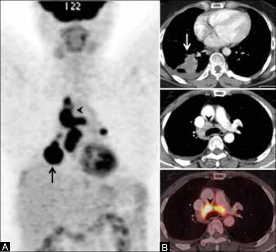
FDG PET in nodal disease false-positive study. Maximum intensity projection (MIP) image shows an FDG-avid primary lung tumor on the right side (arrow, A) and multiple foci of FDG uptake in the mediastinum (arrowhead, A). CT scan shows enhancing, primary tumor (arrow, B). Fused PET/CT image shows FDG concentration in the mediastinal nodes, suggesting metastatic involvement. Mediastinoscopy and biospy revealed tuberculosis
File history
Click on a date/time to view the file as it appeared at that time.
| Date/Time | Thumbnail | Dimensions | User | Comment | |
|---|---|---|---|---|---|
| current | 16:26, 16 February 2018 |  | 539 × 492 (54 KB) | Dildar Hussain (talk | contribs) | FDG PET in nodal disease false-positive study. Maximum intensity projection (MIP) image shows an FDG-avid primary lung tumor on the right side (arrow, A) and multiple foci of FDG uptake in the mediastinum (arrowhead, A). CT scan shows enhancing, primar... |
You cannot overwrite this file.
File usage
The following page uses this file: