File:Hantavirus01.jpeg
Jump to navigation
Jump to search
No higher resolution available.
Hantavirus01.jpeg (700 × 461 pixels, file size: 54 KB, MIME type: image/jpeg)
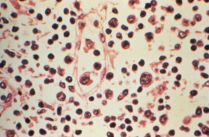
This image reveals some of the cytoarchitectural features seen in a lymph node specimen that had been extracted from a patient suspected of a Hantavirus illness. Note the concentration of lymphohistiocytic infiltrates, almost all cases have expanded paracortical regions, or T-cell regions with immunoblasts, which sometimes extend into the cortex and into the medulla.
File history
Click on a date/time to view the file as it appeared at that time.
| Date/Time | Thumbnail | Dimensions | User | Comment | |
|---|---|---|---|---|---|
| current | 16:16, 11 December 2014 | | 700 × 461 (54 KB) | Jesus Hernandez (talk | contribs) | This image reveals some of the cytoarchitectural features seen in a lymph node specimen that had been extracted from a patient suspected of a Hantavirus illness. Note the concentration of lymphohistiocytic infiltrates, almost all cases have expanded pa... |
You cannot overwrite this file.
File usage
The following file is a duplicate of this file (more details):
The following page uses this file: