File:Group A streptococcus19.jpeg
Group_A_streptococcus19.jpeg (700 × 462 pixels, file size: 22 KB, MIME type: image/jpeg)
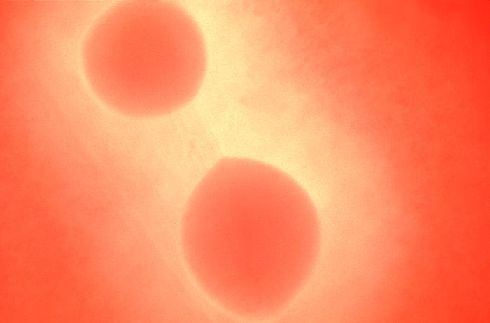
Magnified 100x, this 1977 photograph depicted a Petri dish filled with trypticase soy agar medium containing 5% defibrinated sheep's blood, i.e., blood agar plate (BAP). After having been inoculated by streaking the surface of the BAP with Group A Streptococcus pyogenes (GAS) bacteria, the dish was incubated in a carbon dioxide enriched atmosphere at 35oC for 24 hours. The culture grew bacterial surface colonies. The characteristic color changes, i.e., a colorless region surrounding each colony in which the red blood cells in the blood agar medium had been destroyed, or "hemolyzed", indicated that these bacteria were indeed beta-hemolytic in nature.
File history
Click on a date/time to view the file as it appeared at that time.
| Date/Time | Thumbnail | Dimensions | User | Comment | |
|---|---|---|---|---|---|
| current | 15:00, 2 December 2014 | | 700 × 462 (22 KB) | Jesus Hernandez (talk | contribs) | Magnified 100x, this 1977 photograph depicted a Petri dish filled with trypticase soy agar medium containing 5% defibrinated sheep's blood, i.e., blood agar plate (BAP). After having been inoculated by streaking the surface of the BAP with Group A Stre... |
You cannot overwrite this file.
File usage
The following page uses this file: