File:Group A streptococcus13.jpeg
Group_A_streptococcus13.jpeg (700 × 483 pixels, file size: 24 KB, MIME type: image/jpeg)
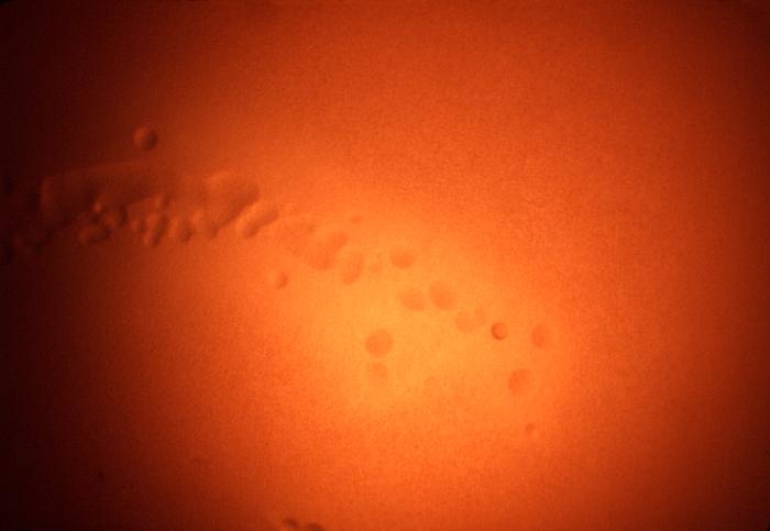
Magnified 100x, this 1977 photograph depicted a Petri dish filled with heart infusion agar medium containing 5% defibrinated rabbit blood, i.e., blood agar plate (BAP). After having been inoculated with a culture of Streptococcus anginosus bacteria, of the Gram-positive viridans group of streptococci (VGS), the BAP was incubated in a carbon dioxide enriched atmosphere at 35oC for 24 hours. In this view, one can see numbers of growing "surface" colonies surrounded by what is known as "wide zone alpha hemolytic" (WZα) color changes. Characteristics of WZα reactivity are described as, "the area immediately adjacent to the colony has some red blood cells (RBCs), but an area outside of that may be completely, or nearly completely, cleared of RBCs. Therefore, there is no reactive zones where "complete" RBC hemolysis has occurred, as is the case in beta-hemolytic reactions, hence the Wide Zone "alpha" terminology.
File history
Click on a date/time to view the file as it appeared at that time.
| Date/Time | Thumbnail | Dimensions | User | Comment | |
|---|---|---|---|---|---|
| current | 14:58, 2 December 2014 | | 700 × 483 (24 KB) | Jesus Hernandez (talk | contribs) | Magnified 100x, this 1977 photograph depicted a Petri dish filled with heart infusion agar medium containing 5% defibrinated rabbit blood, i.e., blood agar plate (BAP). After having been inoculated with a culture of Streptococcus anginosus bacteria, of... |
You cannot overwrite this file.
File usage
The following page uses this file: