File:Group A streptococcus12.jpeg
Group_A_streptococcus12.jpeg (700 × 478 pixels, file size: 35 KB, MIME type: image/jpeg)
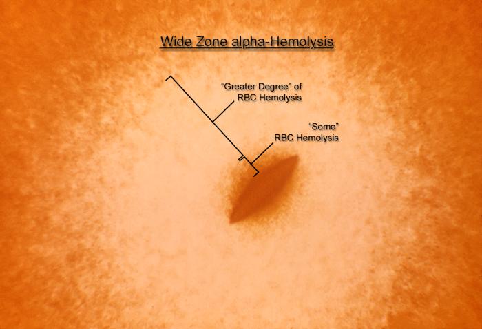
Magnified 100x, this 1977 photograph depicted a Petri dish filled with heart infusion agar medium containing 5% defibrinated rabbit blood, i.e., blood agar plate (BAP). A loop of diluted culture of Streptococcus anginosus was put into the melted agar (50oC) just before the blood was added to the melted agar. The agar was allowed to solidify, and then incubated at 35oC for 24 hours in a normal atmosphere. The culture grew subsurface bacterial colonies, one of which is seen here, surrounded by what is known as "wide zone alpha hemolytic" (WZα) color changes. Characteristics of WZα reactivity are described as, "the area immediately adjacent to the colony has some red blood cells (RBCs), but an area outside of that may be completely, or nearly completely, cleared of RBCs. Therefore, there are no reactive zones where "complete" RBC hemolysis has occurred, as in the case of beta-hemolytic reactions, hence the Wide Zone "alpha" terminology.
File history
Click on a date/time to view the file as it appeared at that time.
| Date/Time | Thumbnail | Dimensions | User | Comment | |
|---|---|---|---|---|---|
| current | 14:57, 2 December 2014 | | 700 × 478 (35 KB) | Jesus Hernandez (talk | contribs) | Magnified 100x, this 1977 photograph depicted a Petri dish filled with heart infusion agar medium containing 5% defibrinated rabbit blood, i.e., blood agar plate (BAP). A loop of diluted culture of Streptococcus anginosus was put into the melted agar (... |
You cannot overwrite this file.
File usage
The following page uses this file: