File:Group A streptococcus11.jpeg
Group_A_streptococcus11.jpeg (700 × 486 pixels, file size: 20 KB, MIME type: image/jpeg)
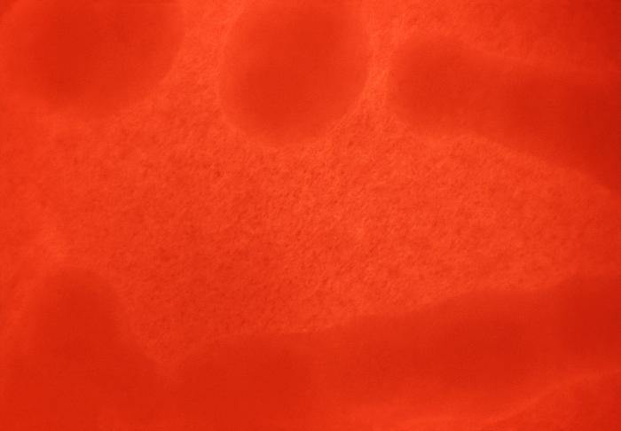
Magnified 100X, this 1977 photograph depicted a Petri dish filled with trypticase soy agar medium containing 5% defibrinated sheep's blood, i.e., blood agar plate (BAP). After having been inoculated by streaking the surface of the BAP with a non-hemolytic group A Streptococcus pyogenes (GAS) bacteria. The BAP was incubated in a carbon dioxide enriched atmosphere at 35oC for 24 hours, and grew bacterial surface colonies with no characteristic color changes surrounding each colony, or in the stabbed areas. Under examination, no red blood cells in the blood agar medium had been altered, or "hemolyzed", indicating that these bacteria were indeed non-hemolytic in nature.
File history
Click on a date/time to view the file as it appeared at that time.
| Date/Time | Thumbnail | Dimensions | User | Comment | |
|---|---|---|---|---|---|
| current | 14:55, 2 December 2014 | | 700 × 486 (20 KB) | Jesus Hernandez (talk | contribs) | Magnified 100X, this 1977 photograph depicted a Petri dish filled with trypticase soy agar medium containing 5% defibrinated sheep's blood, i.e., blood agar plate (BAP). After having been inoculated by streaking the surface of the BAP with a non-hemoly... |
You cannot overwrite this file.
File usage
The following page uses this file: