File:Granulosa cell tumor.jpeg
Granulosa_cell_tumor.jpeg (567 × 567 pixels, file size: 94 KB, MIME type: image/jpeg)
Summary
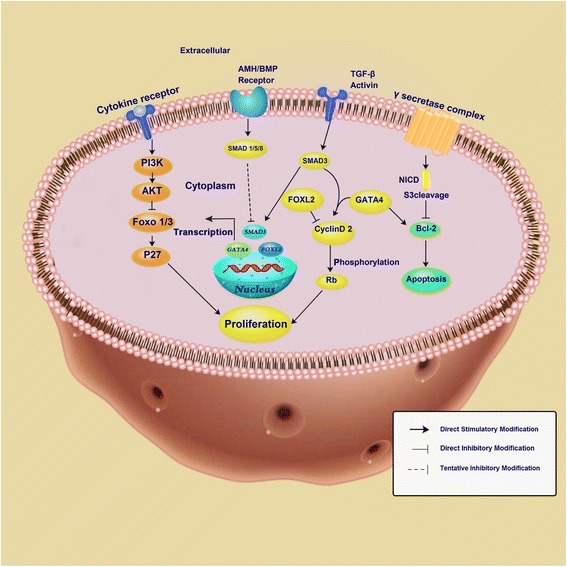
Schematic representation of the cell signaling pathways in GCT development. PI3K, phosphatidylinositol-3-kinase; AKT, serine/threonine kinase; FOXO 1/3, forkhead box O1/3; AMH, anti-Mullerian hormone; BMP, bone morphogenetic protein; SMAD, drosophila mothers against decapentaplegic protein; FOXL2, forkhead transcription factor 2; Rb, retinoblastoma protein; Bcl-2, B-cell lymphoma 2chematic representation of the cell signaling pathways in granulosa cell tumor development. Courtesy: Li J, Bao R, Peng S, Zhang C. The molecular mechanism of ovarian granulosa cell tumors. J Ovarian Res. 2018;11(1):13. Published 2018 Feb 6. doi:10.1186/s13048-018-0384-1
File history
Click on a date/time to view the file as it appeared at that time.
| Date/Time | Thumbnail | Dimensions | User | Comment | |
|---|---|---|---|---|---|
| current | 20:05, 8 March 2019 |  | 567 × 567 (94 KB) | Maneesha Nandimandalam (talk | contribs) | Schematic representation of the cell signaling pathways in GCT development. PI3K, phosphatidylinositol-3-kinase; AKT, serine/threonine kinase; FOXO 1/3, forkhead box O1/3; AMH, anti-Mullerian hormone; BMP, bone morphogenetic protein; SMAD, drosophila m... |
You cannot overwrite this file.
File usage
The following page uses this file: