File:Giardiasis07.jpeg
Jump to navigation
Jump to search
Size of this preview: 408 × 599 pixels. Other resolution: 700 × 1,028 pixels.
Original file (700 × 1,028 pixels, file size: 63 KB, MIME type: image/jpeg)
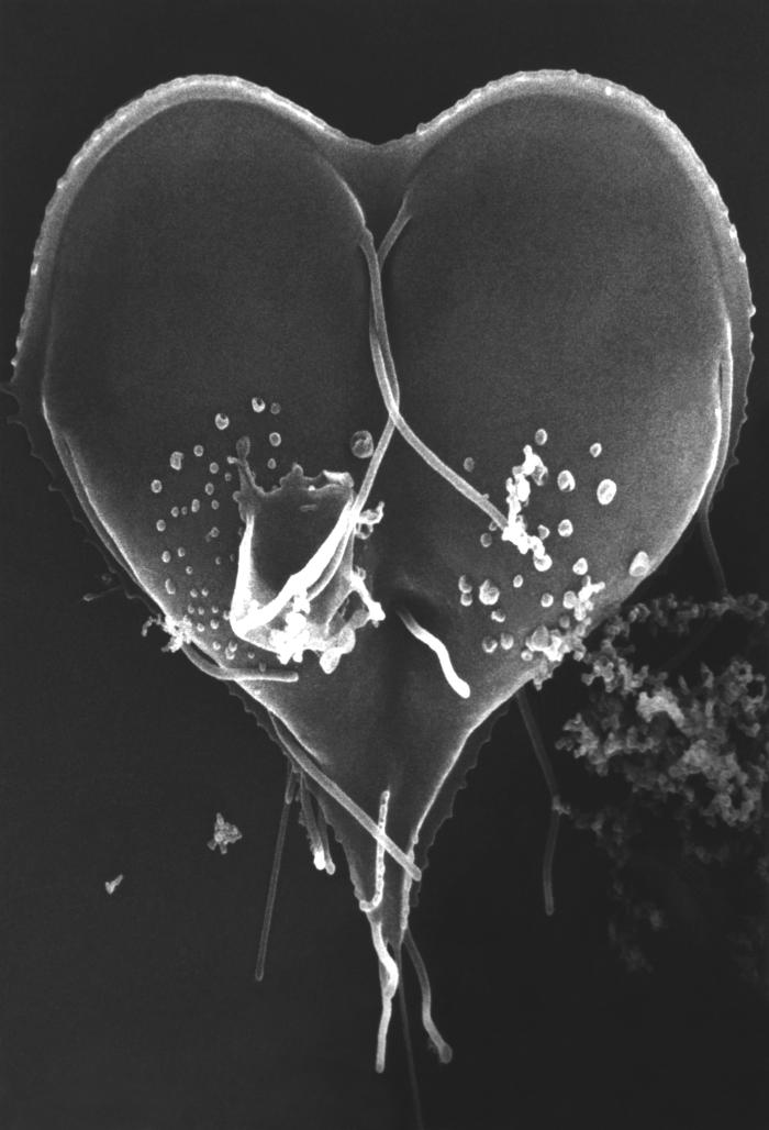
This scanning electron micrograph (SEM) depicted a Giardia lamblia protozoan that was about to become two separate organisms, as it was caught in a late stage of cell division, producing a heart-shaped form. Note the intimate intertwining of two of the organisms’ eight flagella that will facilitate their motility.
File history
Click on a date/time to view the file as it appeared at that time.
| Date/Time | Thumbnail | Dimensions | User | Comment | |
|---|---|---|---|---|---|
| current | 16:20, 10 December 2014 | | 700 × 1,028 (63 KB) | Jesus Hernandez (talk | contribs) | This scanning electron micrograph (SEM) depicted a Giardia lamblia protozoan that was about to become two separate organisms, as it was caught in a late stage of cell division, producing a heart-shaped form. Note the intimate intertwining of two of the... |
You cannot overwrite this file.
File usage
The following page uses this file: