File:Giardiasis04.jpeg
Jump to navigation
Jump to search
No higher resolution available.
Giardiasis04.jpeg (700 × 437 pixels, file size: 81 KB, MIME type: image/jpeg)
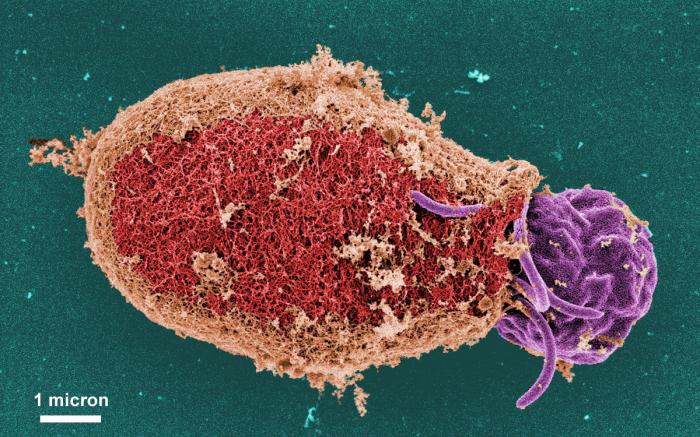
This digitally-colorized scanning electron micrograph (SEM) depicted some of the ultrastructural morphologic details of an oblong-shaped Giardia sp. protozoan cyst, revealing the filamentous nature of the cyst wall. Each cyst-wall filament is approximately 7 to 20 nanometers (nm) thick. Note that this cyst was undergoing "excystation", and was captured at a point in the process where a flagellated trophozoite was beginning to emerge from the right side of the cyst.
File history
Click on a date/time to view the file as it appeared at that time.
| Date/Time | Thumbnail | Dimensions | User | Comment | |
|---|---|---|---|---|---|
| current | 16:18, 10 December 2014 | | 700 × 437 (81 KB) | Jesus Hernandez (talk | contribs) | This digitally-colorized scanning electron micrograph (SEM) depicted some of the ultrastructural morphologic details of an oblong-shaped Giardia sp. protozoan cyst, revealing the filamentous nature of the cyst wall. Each cyst-wall filament is approxima... |
You cannot overwrite this file.
File usage
The following page uses this file: