File:Dracunculiasis10.jpeg
Original file (700 × 926 pixels, file size: 71 KB, MIME type: image/jpeg)
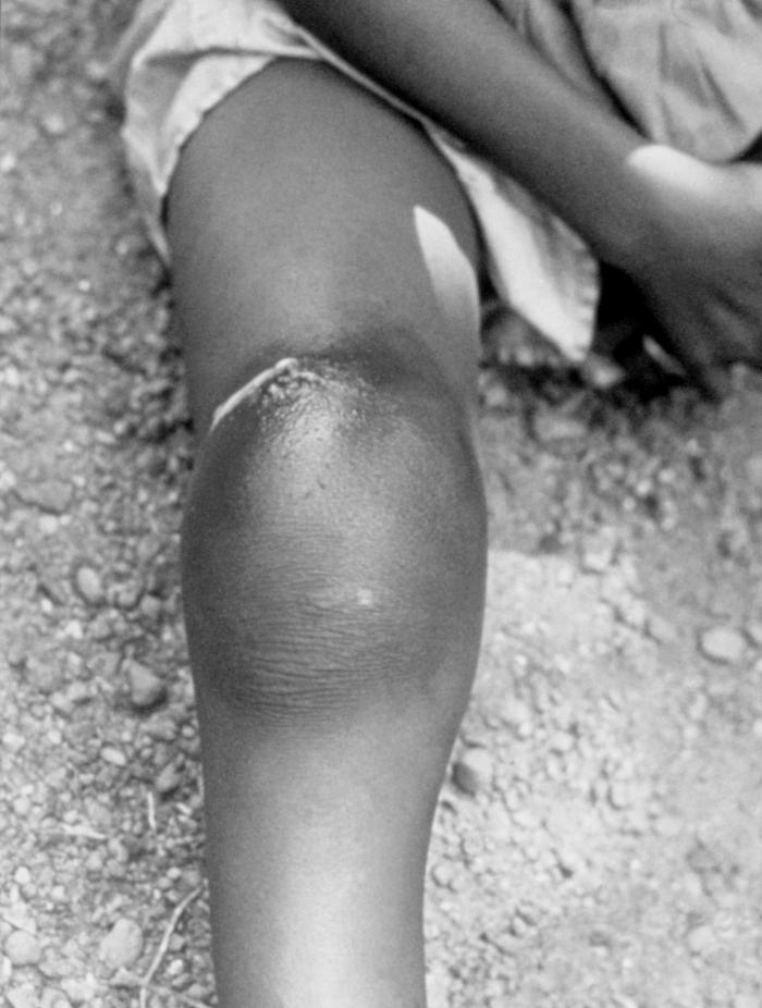
This 1990 image depicted a victim with a severely swollen right knee due to an infection caused by the subcutaneous emergence of an adult female Guinea worm. 10 to 14 months earlier, this child had unintentionally ingested a copepod, i.e., water flea, that was a larval host from a locally Dracunculus medinensis-contaminated water source. As part of the Guinea worm parasite, D. medinensis, life cycle, copepods are carriers of the parasitic larvae, and their ingestion leads to one becoming infected with Guinea worm disease (GWD). Once inside the body, the stomach acid digests the water flea, but not the Guinea worm. These larvae find their way to the small intestine, where they penetrate the wall of the intestine and pass into the body cavity. The female Guinea worm grows to a full size adult 60-100 centimeters (2-3 feet) long, and as wide as a cooked spaghetti noodle. She then migrates to the site where she will emerge, usually from the lower limbs, as was the case here.
File history
Click on a date/time to view the file as it appeared at that time.
| Date/Time | Thumbnail | Dimensions | User | Comment | |
|---|---|---|---|---|---|
| current | 15:02, 8 December 2014 | | 700 × 926 (71 KB) | Jesus Hernandez (talk | contribs) | This 1990 image depicted a victim with a severely swollen right knee due to an infection caused by the subcutaneous emergence of an adult female Guinea worm. 10 to 14 months earlier, this child had unintentionally ingested a copepod, i.e., water flea, ... |
You cannot overwrite this file.
File usage
The following page uses this file: