File:Dracunculiasis04.jpeg
Dracunculiasis04.jpeg (700 × 482 pixels, file size: 49 KB, MIME type: image/jpeg)
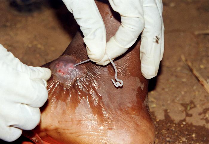
This image depicts the subcutaneous emergence of a female Guinea worm, Dracunculus medinensis, from a sufferer’s lower left leg, just distal to the lateral left knee. The white, spaghetti-like worm is being pulled from the wound by the gloved hand of a health worker. Before the worm emerges, a blister develops on the skin. This blister causes a very painful burning sensation and eventually (within 24 - 72 hours) ruptures. Once the worm emerges from the wound, it can only be pulled out a few centimeters each day and wrapped around a small stick, or piece of gauze. Sometimes the worm can be pulled out completely within a few days, but the process often takes weeks.
File history
Click on a date/time to view the file as it appeared at that time.
| Date/Time | Thumbnail | Dimensions | User | Comment | |
|---|---|---|---|---|---|
| current | 14:53, 8 December 2014 | | 700 × 482 (49 KB) | Jesus Hernandez (talk | contribs) | This image depicts the subcutaneous emergence of a female Guinea worm, Dracunculus medinensis, from a sufferer’s lower left leg, just distal to the lateral left knee. The white, spaghetti-like worm is being pulled from the wound by the gloved hand of... |
You cannot overwrite this file.
File usage
The following page uses this file: