File:Crimean congo hemorrhagic fever02.jpeg
Jump to navigation
Jump to search
No higher resolution available.
Crimean_congo_hemorrhagic_fever02.jpeg (700 × 465 pixels, file size: 89 KB, MIME type: image/jpeg)
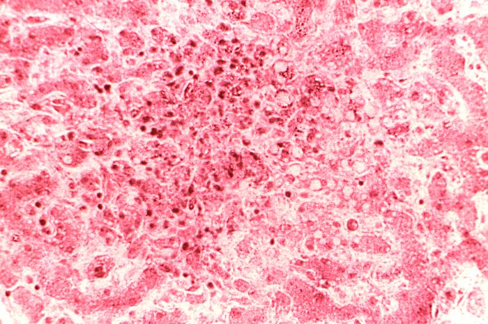
Under a moderate magnification of 280X, this hematoxylin-eosin-stained (H&E) photomicrograph depicts the cytoarchitectural changes found in a liver tissue specimen extracted from a Congo/Crimean hemorrhagic fever patient. This particular view reveals “coagulation necrosis of hepatocytes with an associated perifocal inflammatory reaction. Several cells can be seen to be undergoing fatty degeneration."
File history
Click on a date/time to view the file as it appeared at that time.
| Date/Time | Thumbnail | Dimensions | User | Comment | |
|---|---|---|---|---|---|
| current | 11:43, 5 December 2014 | | 700 × 465 (89 KB) | Jesus Hernandez (talk | contribs) | Under a moderate magnification of 280X, this hematoxylin-eosin-stained (H&E) photomicrograph depicts the cytoarchitectural changes found in a liver tissue specimen extracted from a Congo/Crimean hemorrhagic fever patient. This particular view reveals ... |
You cannot overwrite this file.
File usage
The following page uses this file: