File:Coccidioidomycosis15.jpeg
Jump to navigation
Jump to search
No higher resolution available.
Coccidioidomycosis15.jpeg (700 × 457 pixels, file size: 45 KB, MIME type: image/jpeg)
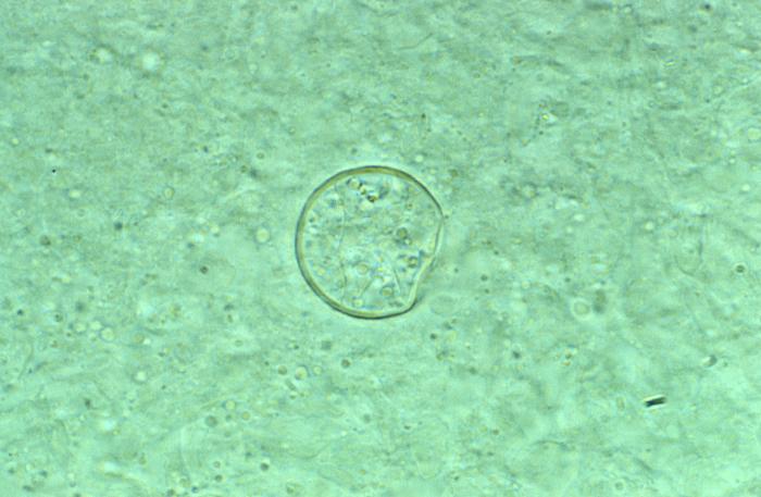
This photomicrograph revealed some of the histopathologic characteristics found within a pus specimen, which was prepared using potassium hudroxide (KOH), and which had been harvested from a skin lesion in a case of cutaneous coccidioidomycosis. In this particular specimen you’ll note the chlamydospore, or spherule of a Coccidioides immitis fungal organism. As the reproductive structure of this, as well as other types of fungi, this spherule is also known as a chlamydoconidium, and contains the organism’s endospores.
File history
Click on a date/time to view the file as it appeared at that time.
| Date/Time | Thumbnail | Dimensions | User | Comment | |
|---|---|---|---|---|---|
| current | 15:25, 4 December 2014 | | 700 × 457 (45 KB) | Jesus Hernandez (talk | contribs) | This photomicrograph revealed some of the histopathologic characteristics found within a pus specimen, which was prepared using potassium hudroxide (KOH), and which had been harvested from a skin lesion in a case of cutaneous coccidioidomycosis. In thi... |
You cannot overwrite this file.
File usage
The following page uses this file: