File:Candidiasis 09.jpeg
Jump to navigation
Jump to search
No higher resolution available.
Candidiasis_09.jpeg (700 × 460 pixels, file size: 58 KB, MIME type: image/jpeg)
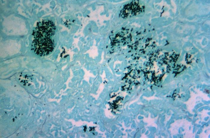
Under a magnification of 125X, this Grocott's (or Gomori’s) methenamine silver stained (GMS) kidney tissue sample revealed the presence of numerous darkly-stained yeast cells of the fungal organism, Candida albicans.
File history
Click on a date/time to view the file as it appeared at that time.
| Date/Time | Thumbnail | Dimensions | User | Comment | |
|---|---|---|---|---|---|
| current | 19:32, 1 December 2014 | | 700 × 460 (58 KB) | Jesus Hernandez (talk | contribs) | Under a magnification of 125X, this Grocott's (or Gomori’s) methenamine silver stained (GMS) kidney tissue sample revealed the presence of numerous darkly-stained yeast cells of the fungal organism, Candida albicans. |
You cannot overwrite this file.
File usage
There are no pages that use this file.