File:Borrelia27.jpeg
Borrelia27.jpeg (700 × 475 pixels, file size: 63 KB, MIME type: image/jpeg)
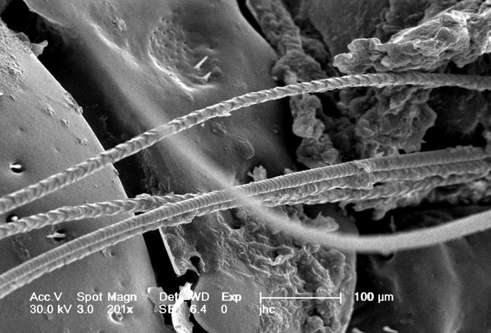
Under a magnification of 201X, this scanning electron micrographic (SEM) image depicted a dorsal view of an unidentified engorged female tick, which had been extracted from the skin of a pet cat while in the process of obtaining its blood meal. Note the presence of some of the cat’s fur, along with some of its skin tissue in which the tick’s gnathosoma were still embedded. See PHIL 9972 and 9973 for additional, less magnified views of this scenario. It is from the “basis capituli” that the two spread “pedipalps”, and hidden skin-piercing hypostome and “chelicerae” emanate. On the dorsal surface of the basis capituli you’ll see two depressed areas known as the “porose areas”, through which secretions produced by dermal glands are released.
File history
Click on a date/time to view the file as it appeared at that time.
| Date/Time | Thumbnail | Dimensions | User | Comment | |
|---|---|---|---|---|---|
| current | 15:13, 26 November 2014 | | 700 × 475 (63 KB) | Jesus Hernandez (talk | contribs) | Under a magnification of 201X, this scanning electron micrographic (SEM) image depicted a dorsal view of an unidentified engorged female tick, which had been extracted from the skin of a pet cat while in the process of obtaining its blood meal. Note th... |
You cannot overwrite this file.
File usage
The following file is a duplicate of this file (more details):
The following page uses this file: