File:Bacterial vaginosis01.jpeg
Jump to navigation
Jump to search
No higher resolution available.
Bacterial_vaginosis01.jpeg (700 × 460 pixels, file size: 44 KB, MIME type: image/jpeg)
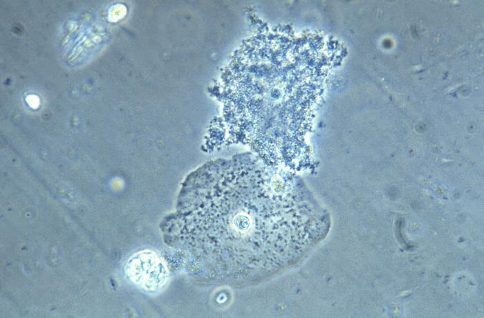
This photomicrograph of a vaginal smear specimen depicts two epithelial cells, a normal cell, and an epithelial cell with its exterior covered by bacteria giving the cell a roughened, stippled appearance known as a “clue cell”.
File history
Click on a date/time to view the file as it appeared at that time.
| Date/Time | Thumbnail | Dimensions | User | Comment | |
|---|---|---|---|---|---|
| current | 21:24, 21 November 2014 | | 700 × 460 (44 KB) | Jesus Hernandez (talk | contribs) | This photomicrograph of a vaginal smear specimen depicts two epithelial cells, a normal cell, and an epithelial cell with its exterior covered by bacteria giving the cell a roughened, stippled appearance known as a “clue cell”. |
You cannot overwrite this file.
File usage
The following file is a duplicate of this file (more details):
The following page uses this file: