File:Aspergillosis04.jpeg
Jump to navigation
Jump to search
Size of this preview: 636 × 600 pixels. Other resolution: 700 × 660 pixels.
Original file (700 × 660 pixels, file size: 44 KB, MIME type: image/jpeg)
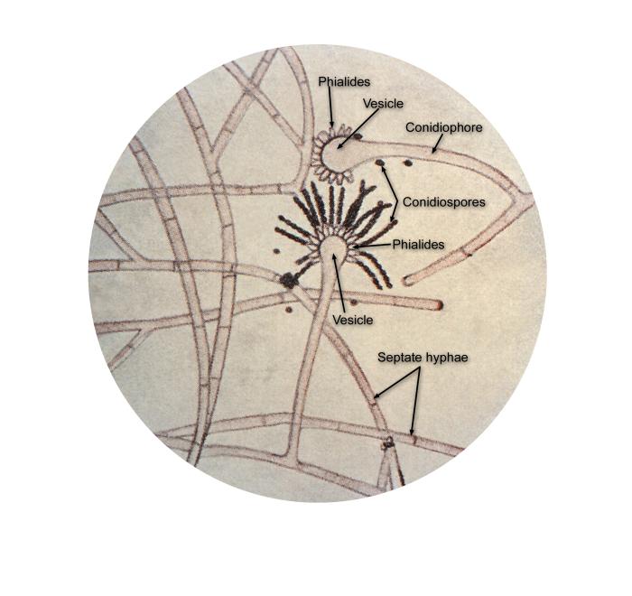
Under a magnification of 125X, this illustration depicts the ultrastructural details found in the common mold, Aspergillus including the organism’s septate hyphae, conidiophores, which support the apparatus responsible for the development of the organism’s asexual conidiospores, i.e., vesicle and phialides.
File history
Click on a date/time to view the file as it appeared at that time.
| Date/Time | Thumbnail | Dimensions | User | Comment | |
|---|---|---|---|---|---|
| current | 15:18, 21 November 2014 | | 700 × 660 (44 KB) | Jesus Hernandez (talk | contribs) | Under a magnification of 125X, this illustration depicts the ultrastructural details found in the common mold, Aspergillus including the organism’s septate hyphae, conidiophores, which support the apparatus responsible for the development of the orga... |
You cannot overwrite this file.
File usage
The following page uses this file: