File:Aspergillosis02.jpeg
Jump to navigation
Jump to search
No higher resolution available.
Aspergillosis02.jpeg (700 × 460 pixels, file size: 29 KB, MIME type: image/jpeg)
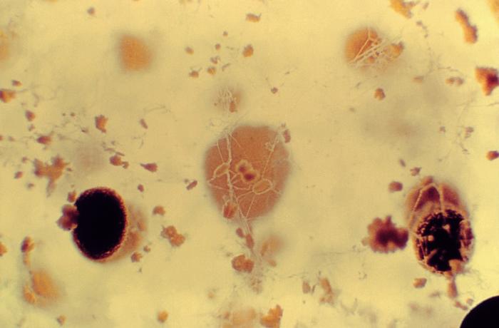
Under a relatively low magnification of 30X, this micrograph of a growing colony of Aspergillus alliaceus revealed some of the ultrastructural characteristics including the presence of sclerotia and conidial heads. Used as a food source during periods of dormancy, sclerotia consist of a hardened mass of the fungus’ own mycelia, and the conidia are the asexually-produced spores, or means of reproduction, which resides atop the conidiophore.
File history
Click on a date/time to view the file as it appeared at that time.
| Date/Time | Thumbnail | Dimensions | User | Comment | |
|---|---|---|---|---|---|
| current | 15:16, 21 November 2014 | | 700 × 460 (29 KB) | Jesus Hernandez (talk | contribs) | Under a relatively low magnification of 30X, this micrograph of a growing colony of Aspergillus alliaceus revealed some of the ultrastructural characteristics including the presence of sclerotia and conidial heads. Used as a food source during periods ... |
You cannot overwrite this file.
File usage
The following page uses this file: