File:Anaplasma phagocytophilum01.jpeg
Jump to navigation
Jump to search
No higher resolution available.
Anaplasma_phagocytophilum01.jpeg (700 × 475 pixels, file size: 63 KB, MIME type: image/jpeg)
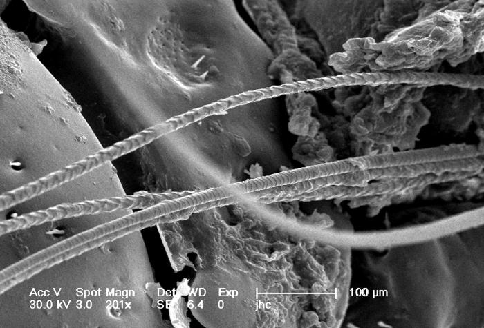
Under a magnification of 201X, this scanning electron micrographic (SEM) image depicted a dorsal view of an unidentified engorged female tick, which had been extracted from the skin of a pet cat while in the process of obtaining its blood meal. Note the presence of some of the cat’s fur, along with some of its skin tissue in which the tick’s gnathosoma were still embedded.
File history
Click on a date/time to view the file as it appeared at that time.
| Date/Time | Thumbnail | Dimensions | User | Comment | |
|---|---|---|---|---|---|
| current | 05:10, 12 December 2014 | | 700 × 475 (63 KB) | Jesus Hernandez (talk | contribs) | Under a magnification of 201X, this scanning electron micrographic (SEM) image depicted a dorsal view of an unidentified engorged female tick, which had been extracted from the skin of a pet cat while in the process of obtaining its blood meal. Note th... |
You cannot overwrite this file.
File usage
The following file is a duplicate of this file (more details):
The following page uses this file: