File:Amebiasis02.jpeg
Amebiasis02.jpeg (700 × 458 pixels, file size: 41 KB, MIME type: image/jpeg)
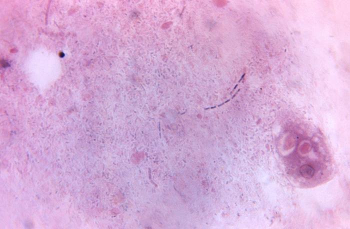
Magnified 675X, this photomicrograph revealed the presence of a parasitic Entamoeba histolytica trophozoite, which contained vacuolated cytoplasm, within which were two red blood cells (RBCs), and a pyknotic body. Entamoeba histolytica/Entamoeba dispar trophozoites have a single nucleus, which have a centrally placed karyosome and uniformly distributed peripheral chromatin. This typical appearance of the nucleus is not always observed as some trophozoites can have nuclei with an eccentric karyosome and unevenly distributed peripheral chromatin. The cytoplasm has a granular or "ground-glass" appearance. E. histolytica/E. dispar trophozoites usually measure 15µm - 20µm (range 10µm - 60µm), tending to be more elongated in diarrheal stool.
File history
Click on a date/time to view the file as it appeared at that time.
| Date/Time | Thumbnail | Dimensions | User | Comment | |
|---|---|---|---|---|---|
| current | 16:38, 20 November 2014 | | 700 × 458 (41 KB) | Jesus Hernandez (talk | contribs) | Magnified 675X, this photomicrograph revealed the presence of a parasitic Entamoeba histolytica trophozoite, which contained vacuolated cytoplasm, within which were two red blood cells (RBCs), and a pyknotic body. Entamoeba histolytica/Entamoeba dispar... |
You cannot overwrite this file.
File usage
The following file is a duplicate of this file (more details):
The following page uses this file: