File:Actinomycosis10.jpeg
Jump to navigation
Jump to search
No higher resolution available.
Actinomycosis10.jpeg (700 × 464 pixels, file size: 31 KB, MIME type: image/jpeg)
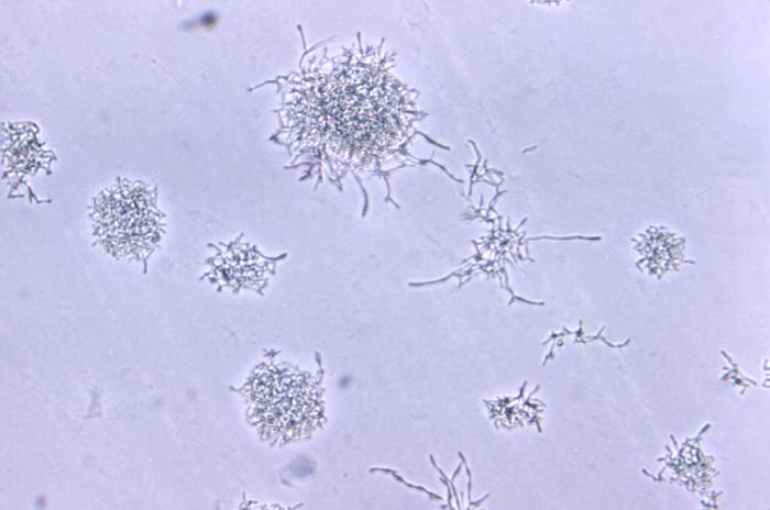
Magnified 800X, this photomicrograph revealed some of the pathologic cytoarchitectural changes seen in a brain abscess tissue specimen due to actinomycosis. One of the primary features, is the presence of a fine network of branching hyphae.
File history
Click on a date/time to view the file as it appeared at that time.
| Date/Time | Thumbnail | Dimensions | User | Comment | |
|---|---|---|---|---|---|
| current | 20:01, 18 November 2014 | | 700 × 464 (31 KB) | Jesus Hernandez (talk | contribs) | Magnified 800X, this photomicrograph revealed some of the pathologic cytoarchitectural changes seen in a brain abscess tissue specimen due to actinomycosis. One of the primary features, is the presence of a fine network of branching hyphae. |
You cannot overwrite this file.
File usage
The following page uses this file: