File:Actinomycosis03.jpeg
Jump to navigation
Jump to search
No higher resolution available.
Actinomycosis03.jpeg (700 × 460 pixels, file size: 51 KB, MIME type: image/jpeg)
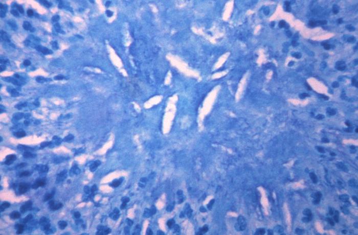
Under a magnification of 200x, and stained using the acid-fast and water-acid staining method, this brain tissue specimen revealed the presence of an actinomycotic sulfur granule, in a rare case of disseminated actinomycosis.
File history
Click on a date/time to view the file as it appeared at that time.
| Date/Time | Thumbnail | Dimensions | User | Comment | |
|---|---|---|---|---|---|
| current | 19:56, 18 November 2014 | | 700 × 460 (51 KB) | Jesus Hernandez (talk | contribs) | Under a magnification of 200x, and stained using the acid-fast and water-acid staining method, this brain tissue specimen revealed the presence of an actinomycotic sulfur granule, in a rare case of disseminated actinomycosis. |
You cannot overwrite this file.
File usage
The following file is a duplicate of this file (more details):
The following page uses this file: