File:Actinomycosis02.jpeg
Jump to navigation
Jump to search
No higher resolution available.
Actinomycosis02.jpeg (700 × 460 pixels, file size: 64 KB, MIME type: image/jpeg)
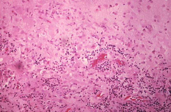
Under a relatively low magnification of 50x, this hematoxylin-eosin (H&E) stained brain tissue specimen, extracted from the occipital cortical region adjacent to an actinomycotic abscess, revealed some of the histopathologic changes associated with a rare case of disseminated actinomycosis.
File history
Click on a date/time to view the file as it appeared at that time.
| Date/Time | Thumbnail | Dimensions | User | Comment | |
|---|---|---|---|---|---|
| current | 17:43, 18 November 2014 | | 700 × 460 (64 KB) | Jesus Hernandez (talk | contribs) | Under a relatively low magnification of 50x, this hematoxylin-eosin (H&E) stained brain tissue specimen, extracted from the occipital cortical region adjacent to an actinomycotic abscess, revealed some of the histopathologic changes associated with a r... |
You cannot overwrite this file.
File usage
The following file is a duplicate of this file (more details):
The following page uses this file: