VIPoma CT: Difference between revisions
Jump to navigation
Jump to search
Madhu Sigdel (talk | contribs) |
Madhu Sigdel (talk | contribs) No edit summary |
||
| Line 5: | Line 5: | ||
On CT scan VIPoma is characterized by hypervascularity with diffuse multiple metastatic nodulation.<ref name="pmid25184777">{{cite journal| author=Apodaca-Torrez FR, Triviño M, Lobo EJ, Goldenberg A, Triviño T| title=Extra-pancreatic vipoma. | journal=Arq Bras Cir Dig | year= 2014 | volume= 27 | issue= 3 | pages= 222-3 | pmid=25184777 | doi= | pmc= | url=http://www.ncbi.nlm.nih.gov/entrez/eutils/elink.fcgi?dbfrom=pubmed&tool=sumsearch.org/cite&retmode=ref&cmd=prlinks&id=25184777 }} </ref> | On CT scan VIPoma is characterized by hypervascularity with diffuse multiple metastatic nodulation.<ref name="pmid25184777">{{cite journal| author=Apodaca-Torrez FR, Triviño M, Lobo EJ, Goldenberg A, Triviño T| title=Extra-pancreatic vipoma. | journal=Arq Bras Cir Dig | year= 2014 | volume= 27 | issue= 3 | pages= 222-3 | pmid=25184777 | doi= | pmc= | url=http://www.ncbi.nlm.nih.gov/entrez/eutils/elink.fcgi?dbfrom=pubmed&tool=sumsearch.org/cite&retmode=ref&cmd=prlinks&id=25184777 }} </ref> | ||
==CT== | ==CT== | ||
On CT scan VIPoma is characterized by hypervascularity with diffuse multiple metastatic nodulation.<ref name="pmid25184777">{{cite journal| author=Apodaca-Torrez FR, Triviño M, Lobo EJ, Goldenberg A, Triviño T| title=Extra-pancreatic vipoma. | journal=Arq Bras Cir Dig | year= 2014 | volume= 27 | issue= 3 | pages= 222-3 | pmid=25184777 | doi= | pmc= | url=http://www.ncbi.nlm.nih.gov/entrez/eutils/elink.fcgi?dbfrom=pubmed&tool=sumsearch.org/cite&retmode=ref&cmd=prlinks&id=25184777 }} </ref> CT scan are highly accurate for tumor localization of primary neuroendocrine pancreatic tumor. Since most of them are more than | On CT scan VIPoma is characterized by hypervascularity with diffuse multiple metastatic nodulation.<ref name="pmid25184777">{{cite journal| author=Apodaca-Torrez FR, Triviño M, Lobo EJ, Goldenberg A, Triviño T| title=Extra-pancreatic vipoma. | journal=Arq Bras Cir Dig | year= 2014 | volume= 27 | issue= 3 | pages= 222-3 | pmid=25184777 | doi= | pmc= | url=http://www.ncbi.nlm.nih.gov/entrez/eutils/elink.fcgi?dbfrom=pubmed&tool=sumsearch.org/cite&retmode=ref&cmd=prlinks&id=25184777 }} </ref> CT scan are highly accurate for tumor localization of primary neuroendocrine pancreatic tumor. Since most of them are more than 2cm in size at the time of presentation. Sensitivity of contrast enhanced CT for VIPoma approaches 100%. | ||
==Gallery== | ==Gallery== | ||
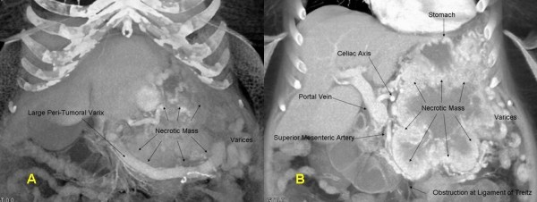
[[Image:CT_VIPoma1.png|900px|thumb|center|(A)3-D CT coronal reconstruction showing the pancreatic VIPoma, a large peri-tumoral varix, and gastric varices. (B) 3-D CT coronal reconstruction depicting relation of pancreatic VIPoma to adjacent vascular structures and stomach. Note presence of varices as well as invasion of tumor into the fourth portion of the duodenum.<ref name="JoyceHong2008">{{cite journal|last1=Joyce|first1=David L|last2=Hong|first2=Kelvin|last3=Fishman|first3=Elliot K|last4=Wisell|first4=Joshua|last5=Pawlik|first5=Timothy M|title=Multi-visceral resection of pancreatic VIPoma in a patient with sinistral portal hypertension|journal=World Journal of Surgical Oncology|volume=6|issue=1|year=2008|pages=80|issn=1477-7819|doi=10.1186/1477-7819-6-80}}</ref>]] | [[Image:CT_VIPoma1.png|900px|thumb|center|(A)3-D CT coronal reconstruction showing the pancreatic VIPoma, a large peri-tumoral varix, and gastric varices. (B) 3-D CT coronal reconstruction depicting relation of pancreatic VIPoma to adjacent vascular structures and stomach. Note presence of varices as well as invasion of tumor into the fourth portion of the duodenum.<ref name="JoyceHong2008">{{cite journal|last1=Joyce|first1=David L|last2=Hong|first2=Kelvin|last3=Fishman|first3=Elliot K|last4=Wisell|first4=Joshua|last5=Pawlik|first5=Timothy M|title=Multi-visceral resection of pancreatic VIPoma in a patient with sinistral portal hypertension|journal=World Journal of Surgical Oncology|volume=6|issue=1|year=2008|pages=80|issn=1477-7819|doi=10.1186/1477-7819-6-80}}</ref>]] | ||
<br clear="left"/> | <br clear="left" /> | ||
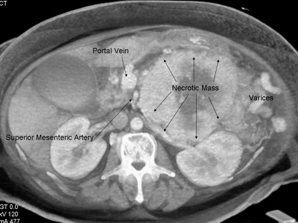
[[Image:CT_VIPoma.jpg|900px|thumb|center|Cross-sectional CT depiction of large necrotic pancreatic VIPoma and its relation to the portal vein and superior mesenteric artery.<ref name="JoyceHong2008">{{cite journal|last1=Joyce|first1=David L|last2=Hong|first2=Kelvin|last3=Fishman|first3=Elliot K|last4=Wisell|first4=Joshua|last5=Pawlik|first5=Timothy M|title=Multi-visceral resection of pancreatic VIPoma in a patient with sinistral portal hypertension|journal=World Journal of Surgical Oncology|volume=6|issue=1|year=2008|pages=80|issn=1477-7819|doi=10.1186/1477-7819-6-80}}</ref>]] | [[Image:CT_VIPoma.jpg|900px|thumb|center|Cross-sectional CT depiction of large necrotic pancreatic VIPoma and its relation to the portal vein and superior mesenteric artery.<ref name="JoyceHong2008">{{cite journal|last1=Joyce|first1=David L|last2=Hong|first2=Kelvin|last3=Fishman|first3=Elliot K|last4=Wisell|first4=Joshua|last5=Pawlik|first5=Timothy M|title=Multi-visceral resection of pancreatic VIPoma in a patient with sinistral portal hypertension|journal=World Journal of Surgical Oncology|volume=6|issue=1|year=2008|pages=80|issn=1477-7819|doi=10.1186/1477-7819-6-80}}</ref>]] | ||
Revision as of 04:16, 13 January 2018
|
VIPoma Microchapters |
|
Diagnosis |
|---|
|
Treatment |
|
Case Studies |
|
VIPoma CT On the Web |
|
American Roentgen Ray Society Images of VIPoma CT |
Editor-In-Chief: C. Michael Gibson, M.S., M.D. [1]Associate Editor(s)-in-Chief: Madhu Sigdel M.B.B.S.[2]Parminder Dhingra, M.D. [3]
Overview
On CT scan VIPoma is characterized by hypervascularity with diffuse multiple metastatic nodulation.[1]
CT
On CT scan VIPoma is characterized by hypervascularity with diffuse multiple metastatic nodulation.[1] CT scan are highly accurate for tumor localization of primary neuroendocrine pancreatic tumor. Since most of them are more than 2cm in size at the time of presentation. Sensitivity of contrast enhanced CT for VIPoma approaches 100%.
Gallery


References
- ↑ 1.0 1.1 Apodaca-Torrez FR, Triviño M, Lobo EJ, Goldenberg A, Triviño T (2014). "Extra-pancreatic vipoma". Arq Bras Cir Dig. 27 (3): 222–3. PMID 25184777.
- ↑ 2.0 2.1 Joyce, David L; Hong, Kelvin; Fishman, Elliot K; Wisell, Joshua; Pawlik, Timothy M (2008). "Multi-visceral resection of pancreatic VIPoma in a patient with sinistral portal hypertension". World Journal of Surgical Oncology. 6 (1): 80. doi:10.1186/1477-7819-6-80. ISSN 1477-7819.