|
|
| (One intermediate revision by the same user not shown) |
| Line 3: |
Line 3: |
| Name = Traumatic diaphragmatic hernia | | | Name = Traumatic diaphragmatic hernia | |
| Image = Diaphragmatic rupture spleen herniation.jpg | | | Image = Diaphragmatic rupture spleen herniation.jpg | |
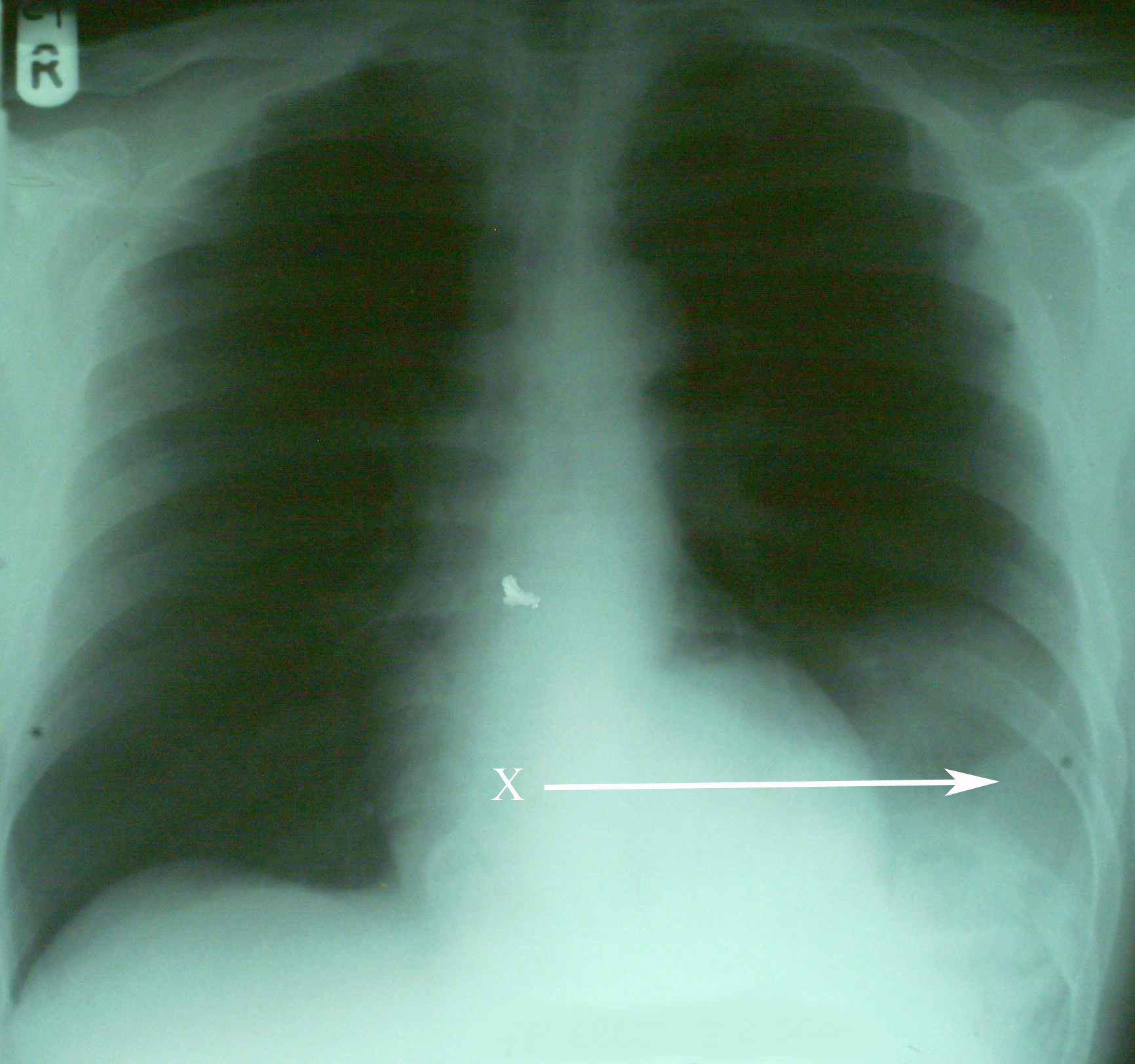
| Caption = An [[X-ray]] showing the [[spleen]] in the left lower portion of the chest cavity (X and arrow) after a diaphragmatic tear<ref name="Hariharan06">{{cite journal |author=Hariharan D, Singhal R, Kinra S, Chilton A |title=Post traumatic intra thoracic spleen presenting with upper GI bleed! A case report |journal=BMC Gastroenterol |volume=6 |issue= |pages=38 |year=2006 |pmid=17132174 |pmc=1687187 |doi=10.1186/1471-230X-6-38 |url=http://www.biomedcentral.com/1471-230X/6/38}}</ref>| | | Caption = An [[X-ray]] showing the [[spleen]] in the left lower portion of the chest cavity (X and arrow) after a diaphragmatic tear| |
| }} | | }} |
| {{Traumatic diaphragmatic hernia}} | | {{Traumatic diaphragmatic hernia}} |
| | '''For patient information, click [[Traumatic diaphragmatic hernia (patient information)|here]]''' |
| | |
| {{CMG}}; '''Associate Editor-in-Chief:''' [[User:AwniShahait|Awni D. Shahait, M.D.]][mailto:awnishahait@yahoo.com], The University of Jordan | | {{CMG}}; '''Associate Editor-in-Chief:''' [[User:AwniShahait|Awni D. Shahait, M.D.]][mailto:awnishahait@yahoo.com], The University of Jordan |
| ==Overview== | | ==[[Traumatic diaphragmatic hernia overview|Overview]]== |
| A '''traumatic diaphragmatic hernia''' is a type of [[diaphragmatic hernia]] which is acquired through an [[abdominal injury]]. This is in contrast to a [[congenital diaphragmatic hernia]], which is present from birth.
| |
| | |
| Diaphragmatic injury accounts for 0.8-1.6% of blunt trauma abdomen. Approximately 4-6% of patients who undergo surgery for trauma have a diaphragmatic injury.<ref name="pmid3738439">{{cite journal |author=Ala-Kulju K, Verkkala K, Ketonen P, Harjola PT |title=Traumatic rupture of the right hemidiaphragm |journal=Scand J Thorac Cardiovasc Surg |volume=20 |issue=2 |pages=109–14 |year=1986 |pmid=3738439 |doi= |url=}}</ref>
| |
|
| |
|
| ==Historical Perspective== | | ==[[Traumatic diaphragmatic hernia historical perspective|Historical Perspective]]== |
| Traumatic diaphragmatic hernia apparently was described by Sennertus, who in 1541 reported an instance of delayed herniation of viscera through an injured diaphragm.<ref name="pmid8526655">{{cite journal |author=Shah R, Sabanathan S, Mearns AJ, Choudhury AK |title=Traumatic rupture of diaphragm |journal=Ann. Thorac. Surg. |volume=60 |issue=5 |pages=1444–9 |year=1995 |month=November |pmid=8526655 |doi=10.1016/0003-4975(95)00629-Y |url=}}</ref> Ambroise Paré, in 1579, described the first case of diaphragmatic rupture diagnosed at autopsy. The first successful diaphragmatic repair was reported by Riolfi in 1886 in a patient with omental prolapse, and Naumann in 1888 repaired the defect with herniated stomach. | |
|
| |
|
| ==Pathophysiology== | | ==[[Traumatic diaphragmatic hernia pathophysiology|Pathophysiology]]== |
| Diaphragmatic injuries are caused either by penetrating or blunt injuries to the abdomen. They are diagnosed immediately as part of multi-organ injury, or present later either with respiratory distress or as intestinal obstruction.<ref name="pmid14799666">{{cite journal |author=CARTER BN, GIUSEFFI J, FELSON B |title=Traumatic diaphragmatic hernia |journal=Am J Roentgenol Radium Ther |volume=65 |issue=1 |pages=56–72 |year=1951 |month=January |pmid=14799666 |doi= |url=}}</ref> The mechanism in blunt injury is explained by shearing of a stretched membrane, avulsion at the point of diaphragmatic attachment, and the sudden force transmission through viscera acting as viscous fluid. Left sided injuries are more often seen. Left-sided rupture occurred in 68.5% of the patients, 24.2% had right-sided rupture, 1.5% had bilateral rupture, 0.9% had pericardial rupture, and 4.9% were unclassified.<ref name="pmid3738439">{{cite journal |author=Ala-Kulju K, Verkkala K, Ketonen P, Harjola PT |title=Traumatic rupture of the right hemidiaphragm |journal=Scand J Thorac Cardiovasc Surg |volume=20 |issue=2 |pages=109–14 |year=1986 |pmid=3738439 |doi= |url=}}</ref> Increased strength of the right hemi-diaphragm, hepatic protection of the right side, under diagnosis of right-sided ruptures, and weakness of the left hemi-diaphragm at points of embryonic fusion all have been proposed to explain the predominance of left sided diaphragmatic injuries.<ref name="pmid3738439">{{cite journal |author=Ala-Kulju K, Verkkala K, Ketonen P, Harjola PT |title=Traumatic rupture of the right hemidiaphragm |journal=Scand J Thorac Cardiovasc Surg |volume=20 |issue=2 |pages=109–14 |year=1986 |pmid=3738439 |doi= |url=}}</ref> Autopsy studies reveals that the incidence of rupture is almost equal on both sides but the greater force needed for the right rupture. A positive pressure gradient of 7-20 cms of H2O between the intraperitoneal and the intra pleural cavities forces the contents into the thorax. With severe blunt trauma the pressures may rise to as high as 100cms of water.
| |
|
| |
|
| It can occur after [[splenectomy]].<ref name="pmid18368327">{{cite journal |author=Tsuboi K, Omura N, Kashiwagi H, Kawasaki N, Suzuki Y, Yanaga K |title=Delayed traumatic diaphragmatic hernia after open splenectomy: report of a case |journal=Surg. Today |volume=38 |issue=4 |pages=352–4 |year=2008 |pmid=18368327 |doi=10.1007/s00595-007-3627-0 |url=http://dx.doi.org/10.1007/s00595-007-3627-0}}</ref>
| | ==[[Traumatic diaphragmatic hernia causes|Causes]]== |
|
| |
|
| Because it can be indicative of severe trauma, it often co-presents with [[pelvic fracture]].<ref name="pmid8257229">{{cite journal |author=Meyers BF, McCabe CJ |title=Traumatic diaphragmatic hernia. Occult marker of serious injury |journal=Ann. Surg. |volume=218 |issue=6 |pages=783–90 |year=1993 |month=December |pmid=8257229 |pmc=1243075 |doi= |url=}}</ref>
| | ==[[Traumatic diaphragmatic hernia differential diagnosis|Differentiating Traumatic Diaphragmatic Hernia from other Diseases]]== |
|
| |
|
| ==Diagnosis== | | ==[[Traumatic diaphragmatic hernia epidemiology and demographics|Epidemiology and Demographics]]== |
| Diaphragmatic ruptures present in two ways. In the acute form, the patient has recently experienced blunt trauma or a penetrating wound to the chest, abdomen, or back. The clinical manifestations are essentially those of the associated injuries, but occasionally, massive herniation of abdominal viscera through the diaphragm causes respiratory insufficiency.
| |
|
| |
| In the chronic form, the diaphragmatic tear is unrecognized at the time of the original injury. Some time later, symptoms appear from herniation of viscera: pain, bowel obstruction, etc. Respiratory symptoms in such cases are rare.
| |
|
| |
|
| The grading of severity has been proposed by Grimes,<ref name="pmid4843862">{{cite journal |author=Grimes OF |title=Traumatic injuries of the diaphragm. Diaphragmatic hernia |journal=Am. J. Surg. |volume=128 |issue=2 |pages=175–81 |year=1974 |month=August |pmid=4843862 |doi= |url=}}</ref> who discussed diaphragmatic rupture in phases: acute, latent and the obstructive phase. The acute presentation is in the patient with poly trauma associated with multiple intra abdominal and chest injuries. The latent phase is when herniation occurs through undetected diaphragmatic ruptures and rents. The obstructive phase is when the loop herniating obstructs and the patient develops distension and strangulation.
| | ==[[Traumatic diaphragmatic hernia risk factors|Risk Factors]]== |
|
| |
|
| ===Investigation=== | | ==[[Traumatic diaphragmatic hernia natural history, complications and prognosis|Natural History, Complications and Prognosis]]== |
| Plain films of the chest may show a radiopaque area and occasionally an air-fluid level if hollow viscera have herniated. If the stomach has entered the chest, the abnormal path of a nasogastric tube may be diagnostic. The collar sign is seen when abdominal contents are seen in the thorax with/without focal constriction. Elevation and distortion of the hemi diaphragm are corroborative signs.<ref name="pmid9460108">{{cite journal |author=Shackleton KL, Stewart ET, Taylor AJ |title=Traumatic diaphragmatic injuries: spectrum of radiographic findings |journal=Radiographics |volume=18 |issue=1 |pages=49–59 |year=1998 |pmid=9460108 |doi= |url=}}</ref>
| |
|
| |
|
| Ultrasonography, CT scan, and MRI may demonstrate the diaphragmatic rent. A CT thorax has a sensitivity of 14-82% and a specificity of 87% and permits direct visualization of the contents and the rupture.Focussed abdominal sonography for trauma(FAST) is now a good aid in diagnosing diaphragmatic hernia.<ref name="pmid15666270">{{cite journal |author=Blaivas M, Brannam L, Hawkins M, Lyon M, Sriram K |title=Bedside emergency ultrasonographic diagnosis of diaphragmatic rupture in blunt abdominal trauma |journal=Am J Emerg Med |volume=22 |issue=7 |pages=601–4 |year=2004 |month=November |pmid=15666270 |doi= |url=}}</ref> Barium study of the colon may show irregular patches of barium in the colon above the diaphragm or a smooth colonic outline if the colon does not contain feces.
| | ==Diagnosis== |
| | | [[Traumatic diaphragmatic hernia history and symptoms|History and Symptoms]] | [[Traumatic diaphragmatic hernia physical examination|Physical Examination]] | [[Traumatic diaphragmatic hernia laboratory findings|Laboratory Findings]] | [[Traumatic diaphragmatic hernia chest x ray|Chest X Ray]] | [[Traumatic diaphragmatic hernia CT|CT]] | [[Traumatic diaphragmatic hernia MRI|MRI]] | [[Traumatic diaphragmatic hernia ultrasound|Ultrasound]] | [[Traumatic diaphragmatic hernia other imaging findings|Other Imaging Findings]] | [[Traumatic diaphragmatic hernia other diagnostic studies|Other Diagnostic Studies]] |
| ==Differential Diagnosis==
| |
| Traumatic rupture of the diaphragm must be differentiated from atelectasis, space-consuming tumors of the lower pleural space, pleural effusion, and intestinal obstruction due to other causes. | |
| | |
| ==Complications==
| |
| Hemorrhage and obstruction may occur. If herniation is massive, progressive cardiorespiratory insufficiency may threaten life. The most severe complication is strangulating obstruction of the herniated viscera.
| |
|
| |
|
| ==Treatment== | | ==Treatment== |
| For acute ruptures, a transabdominal (most commonly) or transthoracic route is used depending on the procedure required to treat ancillary injuries. When the diaphragmatic tear is the only injury, it is usually fixed by laparotomy. Chronic injuries can be repaired by either approach. Asymptomatic tears of the diaphragm with herniated viscera should be repaired, because the risk of strangulating obstruction is high.
| | [[Traumatic diaphragmatic hernia medical therapy|Medical Therapy]] | [[Traumatic diaphragmatic hernia surgery|Surgery]] | [[Traumatic diaphragmatic hernia primary prevention|Primary Prevention]] | [[Traumatic diaphragmatic hernia secondary prevention|Secondary Prevention]] | [[Traumatic diaphragmatic hernia cost-effectiveness of therapy|Cost-Effectiveness of Therapy]] | [[Traumatic diaphragmatic hernia future or investigational therapies|Future or Investigational Therapies]] |
|
| |
|
| ==Prognosis== | | == Case Studies == |
| Surgical repair of the rent in the diaphragm is curative, and the prognosis is excellent. The diaphragm supports sutures well, so that recurrence is practically unknown.
| | [[Traumatic diaphragmatic hernia case study one|Case #1]] |
|
| |
|
| ==Related Chapters== | | ==Related Chapters== |
| *[[Diaphragmatic rupture]] | | *[[Diaphragmatic rupture]] |
|
| |
| ==References==
| |
| {{reflist|2}}
| |
|
| |
|
| |
|
| [[Category:Emergency medicine]] | | [[Category:Emergency medicine]] |
| [[Category:Pulmonology]] | | [[Category:Pulmonology]] |
| | [[Category:Disease]] |
| {{WH}} | | {{WH}} |
| {{WikiDoc Sources}} | | {{WikiDoc Sources}} |
