Thyroid adenoma pathophysiology
|
Thyroid adenoma Microchapters |
|
Diagnosis |
|---|
|
Treatment |
|
Case Studies |
|
Thyroid adenoma pathophysiology On the Web |
|
American Roentgen Ray Society Images of Thyroid adenoma pathophysiology |
|
Risk calculators and risk factors for Thyroid adenoma pathophysiology |
Editor-In-Chief: C. Michael Gibson, M.S., M.D. [3]; Associate Editor(s)-in-Chief: Ammu Susheela, M.D. [4]
Overview
Pathogenesis
A thyroid adenoma may be clinically silent, or it may be a "functional" tumor, producing excessive thyroid hormone. In this case, it may result in symptomatic hyperthyroidism, and may be referred to as a toxic thyroid adenoma. Careful pathological examination may be necessary to distinguish a thyroid adenoma from a minimally invasive follicular thyroid carcinoma.
Genetics
Associated Conditions
Gross Pathology
Thyroid follicular adenoma ranges in diameter from 3 cm on an average, but sometimes is larger (up to 10 cm) or smaller. The typical thyroid adenoma is solitary, spherical and encapsulated lesion that is well demarcated from the surrounding parenchyma. The color ranges from gray-white to red-brown, depending upon
- the cellularity of the adenoma
- the colloid content.
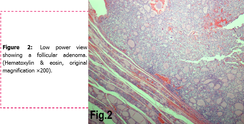
Microscopic Pathology
Areas of hemorrhage, fibrosis, calcification, and cystic change, similar to what is found in multinodular goiters, are common in thyroid (follicular) adenoma, particularly in larger lesions.
-
ADRENAL GLAND: BILATERAL PHEOCHROMOCYTOMA Cross section of bilateral pheochromocytomas from a 30-year-old man with MEN syndrome type IIa. The right adrenal tumor weighed 168 g and the left 220 g. Note the distinct multinodular, multicentric pattern of growth on both sides
-
![Histology of the encapsulated tumor. (a) Photomicrograph showing an encapsulated tumor composed of cells arranged in microfollicular, glandular and trabecular patterns (hematoxylin and eosin; 100×). (b) High power photomicrograph showing the microfollicles containing inspissated colloid resembling hyaline globules and separated by eosinophilic extracellular hyaline material. (hematoxylin and eosin; 100×).[1]](/images/d/dc/Micropathology.jpg)
Histology of the encapsulated tumor. (a) Photomicrograph showing an encapsulated tumor composed of cells arranged in microfollicular, glandular and trabecular patterns (hematoxylin and eosin; 100×). (b) High power photomicrograph showing the microfollicles containing inspissated colloid resembling hyaline globules and separated by eosinophilic extracellular hyaline material. (hematoxylin and eosin; 100×).[1]
-
![(a) Cytoplasmic and membrane positivity for cytokeratin 7 immunostain (400×); (b)negativity for cytokeratin 20 immunostain (400×); (c) nuclear positivity for retinoblastoma immunostain (400×); and (d) extracellular positivity for collagen type IV immunostain (400×).[1]](/images/d/d1/Microfollicular_stain.jpg)
(a) Cytoplasmic and membrane positivity for cytokeratin 7 immunostain (400×); (b)negativity for cytokeratin 20 immunostain (400×); (c) nuclear positivity for retinoblastoma immunostain (400×); and (d) extracellular positivity for collagen type IV immunostain (400×).[1]

![(a) Cytoplasmic and membrane positivity for cytokeratin 7 immunostain (400×); (b)negativity for cytokeratin 20 immunostain (400×); (c) nuclear positivity for retinoblastoma immunostain (400×); and (d) extracellular positivity for collagen type IV immunostain (400×).[1]](/index.php/File:Microfollicular_stain.jpg)
Reference
- ↑ 1.0 1.1 Image courtesy of Dr Frank Gaillard. [1] (original file[2]).Creative Commons BY-SA-NC