Thyroid adenoma biopsy: Difference between revisions
No edit summary |
|||
| Line 3: | Line 3: | ||
{{CMG}}; {{AE}} {{Ammu}} | {{CMG}}; {{AE}} {{Ammu}} | ||
==Overview== | ==Overview== | ||
Fine needle aspiration biopsy may be helpful in [[diagnosis]] of thyroid adenoma. Findings on fine needle aspiration biopsy suggestive of thyroid adenoma include cystic changes, [[fibrosis]], and areas of [[hemorrhage]]. | |||
[[Needle aspiration biopsy|Fine needle aspiration biopsy]] may be helpful in [[diagnosis]] of thyroid adenoma. Findings on fine needle aspiration biopsy suggestive of thyroid adenoma include cystic changes, [[fibrosis]], and areas of [[hemorrhage]]. | |||
==Biopsy== | ==Biopsy== | ||
* One approach used to determine type of adenoma is fine needle biopsy (FNB), which some have described as the most cost-effective, sensitive, and accurate test.<ref> | * One approach used to determine type of adenoma is fine needle biopsy (FNB), which some have described as the most cost-effective, sensitive, and accurate test.<ref> | ||
| Line 31: | Line 33: | ||
}} | }} | ||
</ref> | </ref> | ||
* Fine needle biopsy | * [[Needle aspiration biopsy|Fine needle aspiration biopsy]] or ultrasound-guided [[Needle aspiration biopsy|Fine needle aspiration biopsy]] usually yields sufficient thyroid cells to assess the [[pathology]], although in some cases, the suspected nodule may need to be removed surgically for pathological examination. | ||
===Key Biopsy Findings in Thyroid adenoma=== | ===Key Biopsy Findings in Thyroid adenoma=== | ||
* Areas of [[hemorrhage]], [[fibrosis]], [[calcification]], and cystic change are common in thyroid (follicular) adenoma, particularly in larger lesions. | * Areas of [[hemorrhage]], [[fibrosis]], [[calcification]], and cystic change are common in thyroid (follicular) adenoma, particularly in larger lesions. | ||
* Encapsulated [[tumor]]s do not have any evidence of infiltration. | * Encapsulated [[tumor]]s do not have any evidence of infiltration. | ||
* Colloid nodules are distinguished by an apparently gelatinous mass of [[colloid]] both surrounding and contained within [[follicular cell]]s. Colloid nodules are not surrounded by a [[fibrous capsule]] of compressed tissue. However, they are surrounded by flattened [[epithelium|epithelial]] cells.<ref>{{cite web |url=http://rcpa.tv/parts/educational/anatomical/Dr_Alpha_Tsui/Thyroid_cytology.pdf |title=Thyroid cytology |author=Dr. Alpha Tsui |date=10 October 2010 |publisher=thyroidmanager.org |accessdate=26 September 2011}}</ref> Both the number of cells and the type of colloid may vary considerably.<ref>{{cite web |url=http://www.thyroidmanager.org/chapter%206d/fnabiopsy-frame.htm |title=Fine-Needle Aspiration Biopsy of the Thyroid Gland, Chapter 6d. |author=Diana S. Dean, M.D. Hossein Gharib, M.D. |date=10 October 2010 |publisher=thyroidmanager.org |accessdate=26 September 2011}}</ref> | * Colloid nodules are distinguished by an apparently gelatinous mass of [[colloid]] both surrounding and contained within [[follicular cell]]s. Colloid nodules are not surrounded by a [[fibrous capsule]] of compressed tissue. However, they are surrounded by flattened [[epithelium|epithelial]] cells.<ref>{{cite web |url=http://rcpa.tv/parts/educational/anatomical/Dr_Alpha_Tsui/Thyroid_cytology.pdf |title=Thyroid cytology |author=Dr. Alpha Tsui |date=10 October 2010 |publisher=thyroidmanager.org |accessdate=26 September 2011}}</ref> Both the number of cells and the type of colloid may vary considerably.<ref>{{cite web |url=http://www.thyroidmanager.org/chapter%206d/fnabiopsy-frame.htm |title=Fine-Needle Aspiration Biopsy of the Thyroid Gland, Chapter 6d. |author=Diana S. Dean, M.D. Hossein Gharib, M.D. |date=10 October 2010 |publisher=thyroidmanager.org |accessdate=26 September 2011}}</ref> | ||
===Biopsy Exams of Thyroid adenoma=== | ===Biopsy Exams of Thyroid adenoma=== | ||
<gallery> | <gallery> | ||
Revision as of 21:25, 12 October 2015
|
Thyroid adenoma Microchapters |
|
Diagnosis |
|---|
|
Treatment |
|
Case Studies |
|
Thyroid adenoma biopsy On the Web |
|
American Roentgen Ray Society Images of Thyroid adenoma biopsy |
|
Risk calculators and risk factors for Thyroid adenoma biopsy |
Editor-In-Chief: C. Michael Gibson, M.S., M.D. [3]; Associate Editor(s)-in-Chief: Ammu Susheela, M.D. [4]
Overview
Fine needle aspiration biopsy may be helpful in diagnosis of thyroid adenoma. Findings on fine needle aspiration biopsy suggestive of thyroid adenoma include cystic changes, fibrosis, and areas of hemorrhage.
Biopsy
- One approach used to determine type of adenoma is fine needle biopsy (FNB), which some have described as the most cost-effective, sensitive, and accurate test.[1][2]
- Fine needle aspiration biopsy or ultrasound-guided Fine needle aspiration biopsy usually yields sufficient thyroid cells to assess the pathology, although in some cases, the suspected nodule may need to be removed surgically for pathological examination.
Key Biopsy Findings in Thyroid adenoma
- Areas of hemorrhage, fibrosis, calcification, and cystic change are common in thyroid (follicular) adenoma, particularly in larger lesions.
- Encapsulated tumors do not have any evidence of infiltration.
- Colloid nodules are distinguished by an apparently gelatinous mass of colloid both surrounding and contained within follicular cells. Colloid nodules are not surrounded by a fibrous capsule of compressed tissue. However, they are surrounded by flattened epithelial cells.[3] Both the number of cells and the type of colloid may vary considerably.[4]
Biopsy Exams of Thyroid adenoma
-
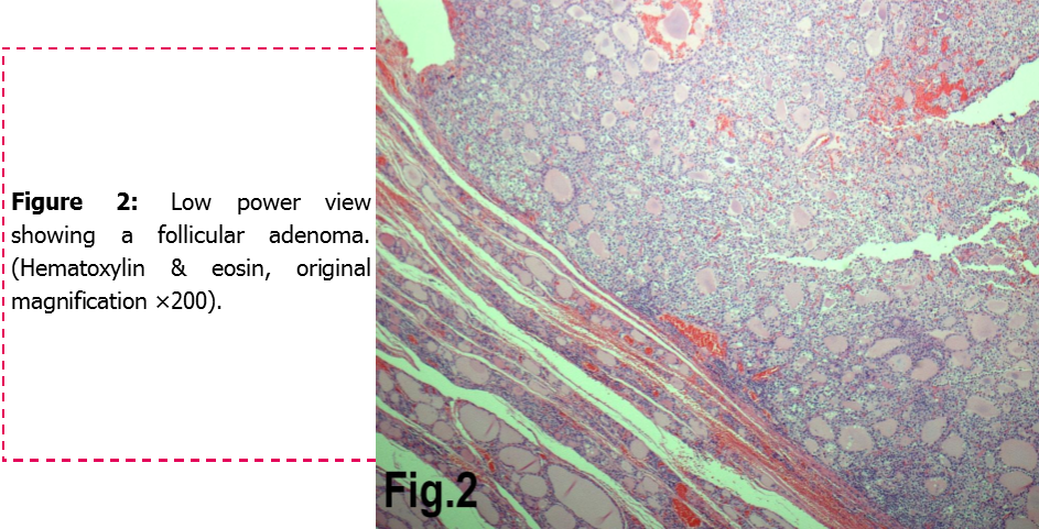
Follicular adenoma of the thyroid gland
-
![Histology of the encapsulated tumor. (a) Photomicrograph showing an encapsulated tumor composed of cells arranged in microfollicular, glandular and trabecular patterns (hematoxylin and eosin; 100×). (b) High power photomicrograph showing the microfollicles containing inspissated colloid resembling hyaline globules and separated by eosinophilic extracellular hyaline material. (hematoxylin and eosin; 100×).[5]](/images/d/dc/Micropathology.jpg)
Histology of the encapsulated tumor. (a) Photomicrograph showing an encapsulated tumor composed of cells arranged in microfollicular, glandular and trabecular patterns (hematoxylin and eosin; 100×). (b) High power photomicrograph showing the microfollicles containing inspissated colloid resembling hyaline globules and separated by eosinophilic extracellular hyaline material. (hematoxylin and eosin; 100×).[5]

References
- ↑ Hamberger, B (1982). "Fine-needle aspiration biopsy of thyroid nodules. Impact on thyroid practice and cost of care". Am J Med. 73 (3): 381–384. doi:10.1016/0002-9343(82)90731-8. PMID 7124765.
- ↑ Mazzaferri (1993). "Management of a Solitary Thyroid Nodule". N Engl J Med. 328 (8): 553–9. doi:10.1056/NEJM199302253280807. PMID 8426623.
- ↑ Dr. Alpha Tsui (10 October 2010). "Thyroid cytology" (PDF). thyroidmanager.org. Retrieved 26 September 2011.
- ↑ Diana S. Dean, M.D. Hossein Gharib, M.D. (10 October 2010). "Fine-Needle Aspiration Biopsy of the Thyroid Gland, Chapter 6d". thyroidmanager.org. Retrieved 26 September 2011.
- ↑ Image courtesy of Dr Frank Gaillard. [1] (original file[2]).Creative Commons BY-SA-NC