Spondyloarthropathy x ray: Difference between revisions
Jump to navigation
Jump to search
(→X-Ray) |
(→X-Ray) |
||
| Line 1: | Line 1: | ||
== X-Ray == | == X-Ray == | ||
{{Spondyloarthropathy}} | |||
* A stepping stone in evaluating of patients with SpA is sacroiliac X-ray. Sacroiliac involvement is a necessary for diagnosis of the disease, which demonstrate bilateral inflammatory condition eventuated in multiple bone erosions and sclerosis of joints. | * A stepping stone in evaluating of patients with SpA is sacroiliac X-ray. Sacroiliac involvement is a necessary for diagnosis of the disease, which demonstrate bilateral inflammatory condition eventuated in multiple bone erosions and sclerosis of joints. | ||
Latest revision as of 22:02, 29 August 2018
X-Ray
|
Spondyloarthropathy Microchapters |
|
Diagnosis |
|---|
|
Treatment |
|
Case Studies |
|
Spondyloarthropathy x ray On the Web |
|
American Roentgen Ray Society Images of Spondyloarthropathy x ray |
|
Risk calculators and risk factors for Spondyloarthropathy x ray |
- A stepping stone in evaluating of patients with SpA is sacroiliac X-ray. Sacroiliac involvement is a necessary for diagnosis of the disease, which demonstrate bilateral inflammatory condition eventuated in multiple bone erosions and sclerosis of joints.
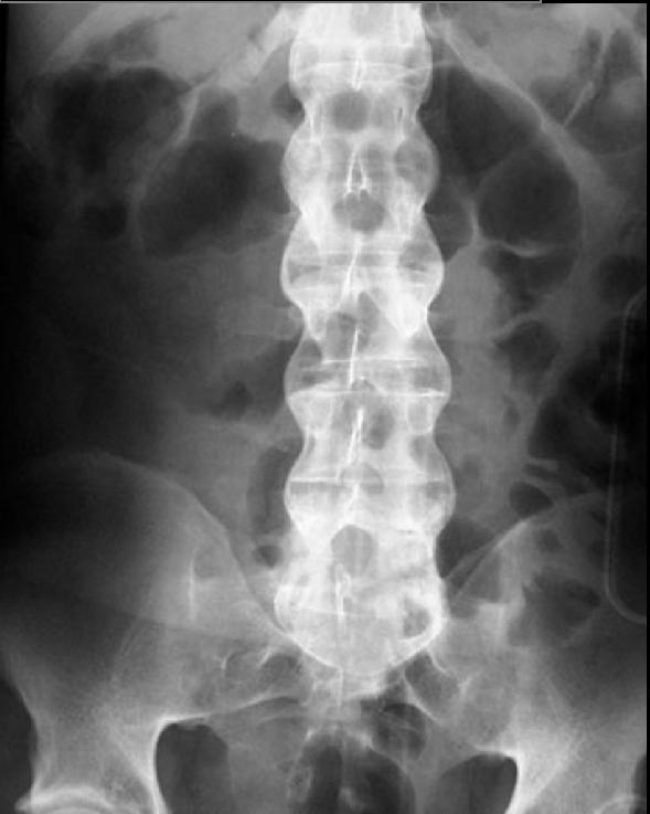
- AS in radiographic studies appear as bilateral, symmetric, and gradually progressive throughout years of disease. At the onset of radiographic signs, subchondral bone plate blurred and afterwards progress to erosions of the margins of the sacroiliac (SI) joint to sclerosis.
- The lower part of the SI joint involve earlier in the disease progression.
- AS enthesitis sign in radiographic studies occurred due to inflammation of annulus fibrosus.
- The first sign is cubic vertebral bodies.

- Annulus fibrosus ossification eventuate in the radiographic appliance of syndesmophwytes. In the progression of the disease over time it can leads to bamboo spine.
- patients with AS are vulnerable to any spinal trauma, and any even low power traumas must be evaluated. Due to ossification of enthuses, ligaments, and other artifacts may obscure the fracture.