Gallery of new files
Jump to navigation
Jump to search
This special page shows the last uploaded files.
-
-
-
-

-
-
-
-
-
-
-

-
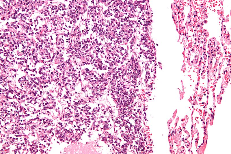
 Medulloblastoma Areas of geographic necrosis.jpg Haytham Allaham
14:14, 5 October 2015
120 × 89; 5 KB
-

-
-
-
-
-

-
-
-
-
-
-

-

-

-

-

-

-

-

-

-

-
-

-

-

-
-

-
-
-
-
-
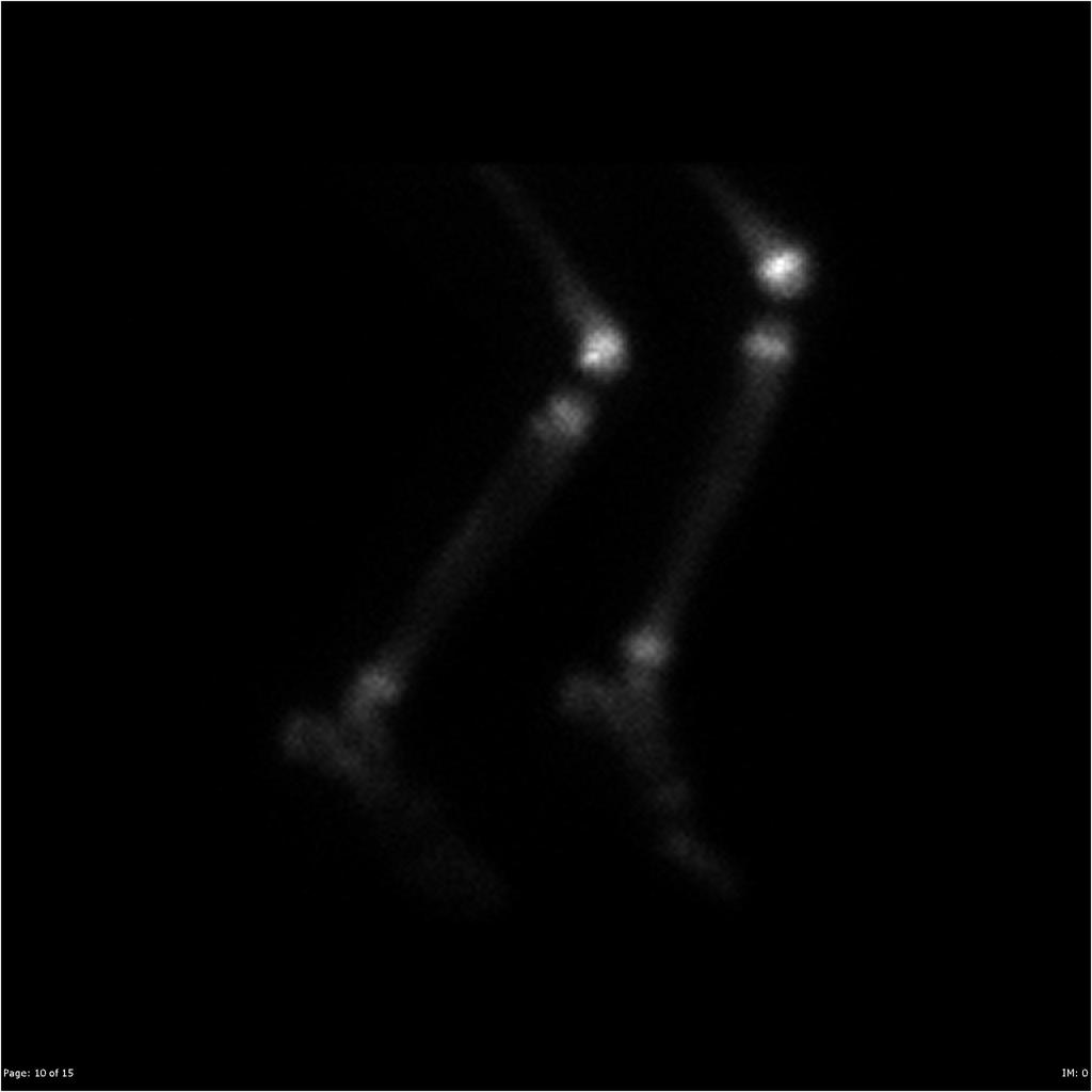
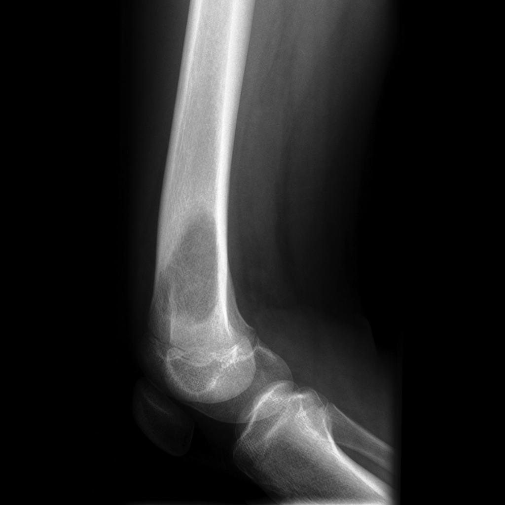
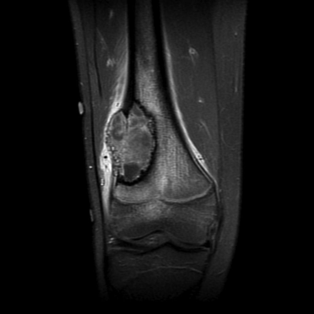 Osteosarcoma-distal-femur MRI T1c.jpg Suveenkrishna Pothuru
15:24, 1 October 2015
1,024 × 1,024; 50 KB
-
-
-
-

-

-

-
-
-
-
-
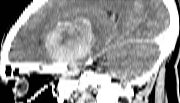
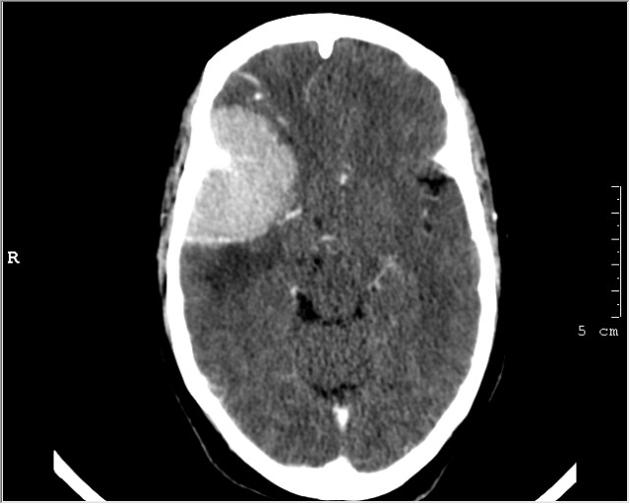
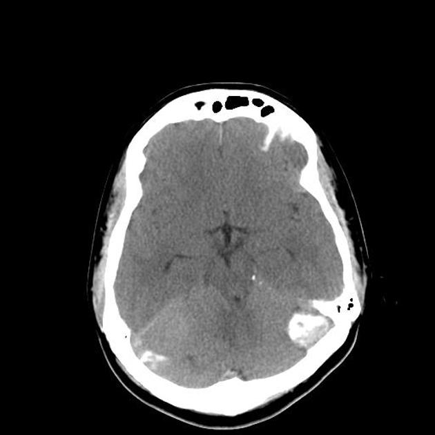
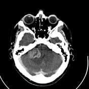
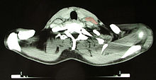
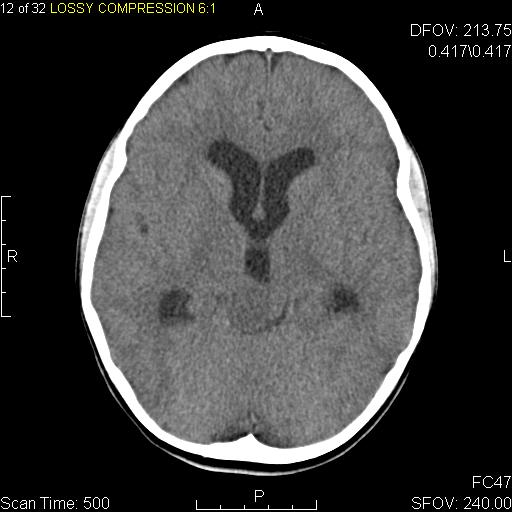
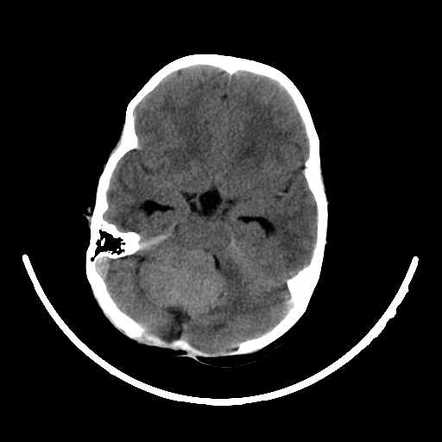 CT noncontrast medulloblastoma Dr Mohammad Taghi Niknejad.jpg Haytham Allaham
12:00, 1 October 2015
442 × 442; 13 KB
-
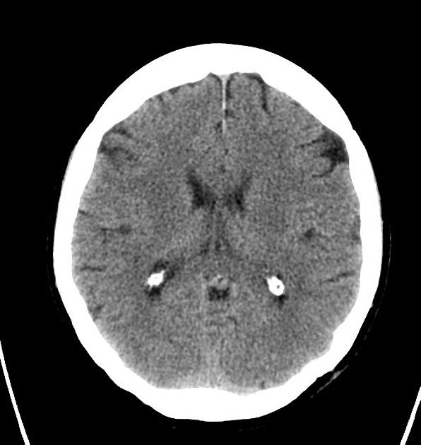
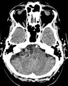
 CT scan noncontrast medulloblastoma Dr Nasir Siddiqui.jpg Haytham Allaham
11:58, 1 October 2015
442 × 442; 21 KB
-
-
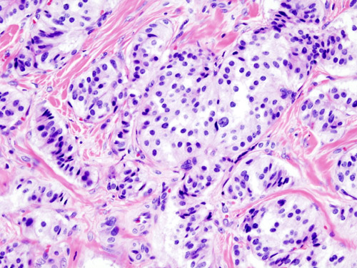
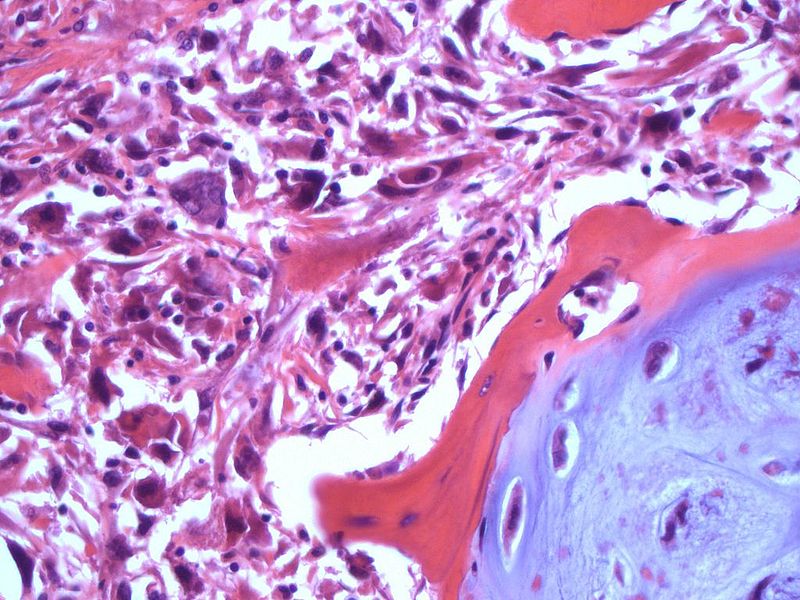
 Osteosarcoma-conventional-histology (1).jpg Suveenkrishna Pothuru
20:38, 30 September 2015
1,024 × 1,024; 151 KB
-
-
-
-
-
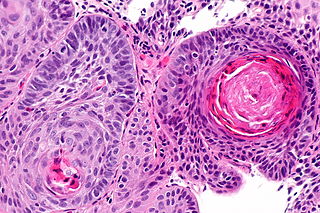
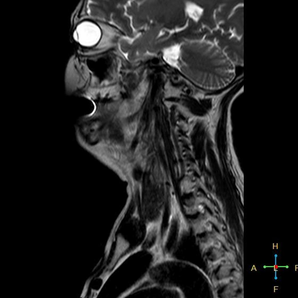
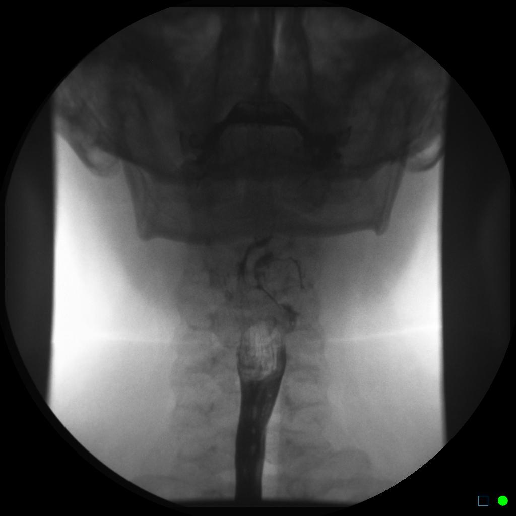
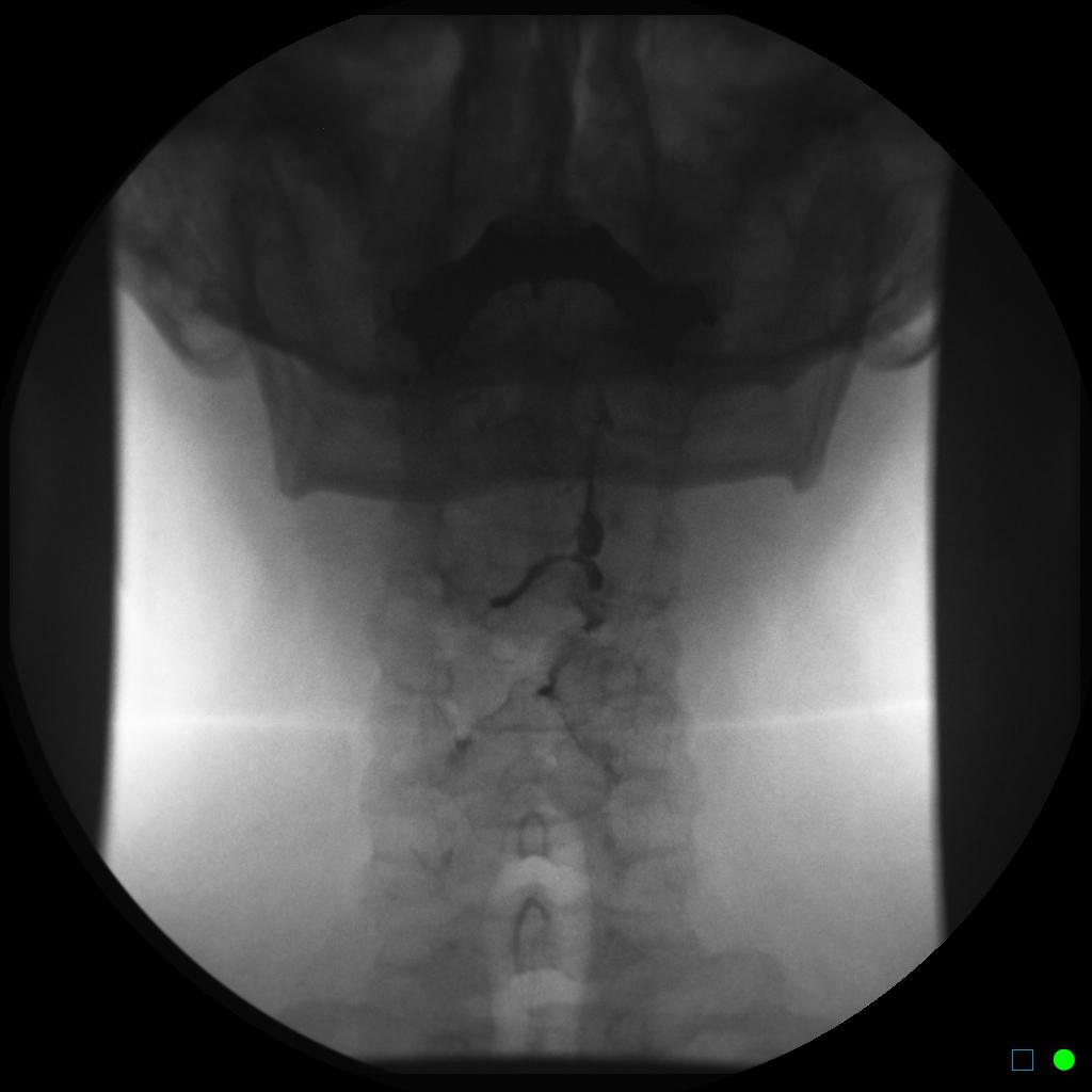
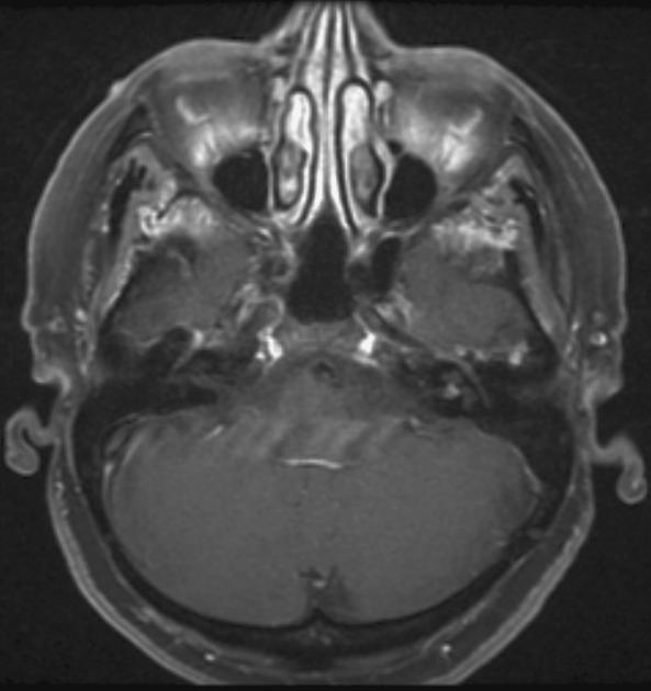
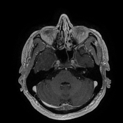
 Left-piriform-fossa-mass-likely-scc (3).jpg Faizan Sheraz
17:12, 30 September 2015
1,020 × 1,024; 58 KB
-
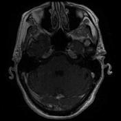
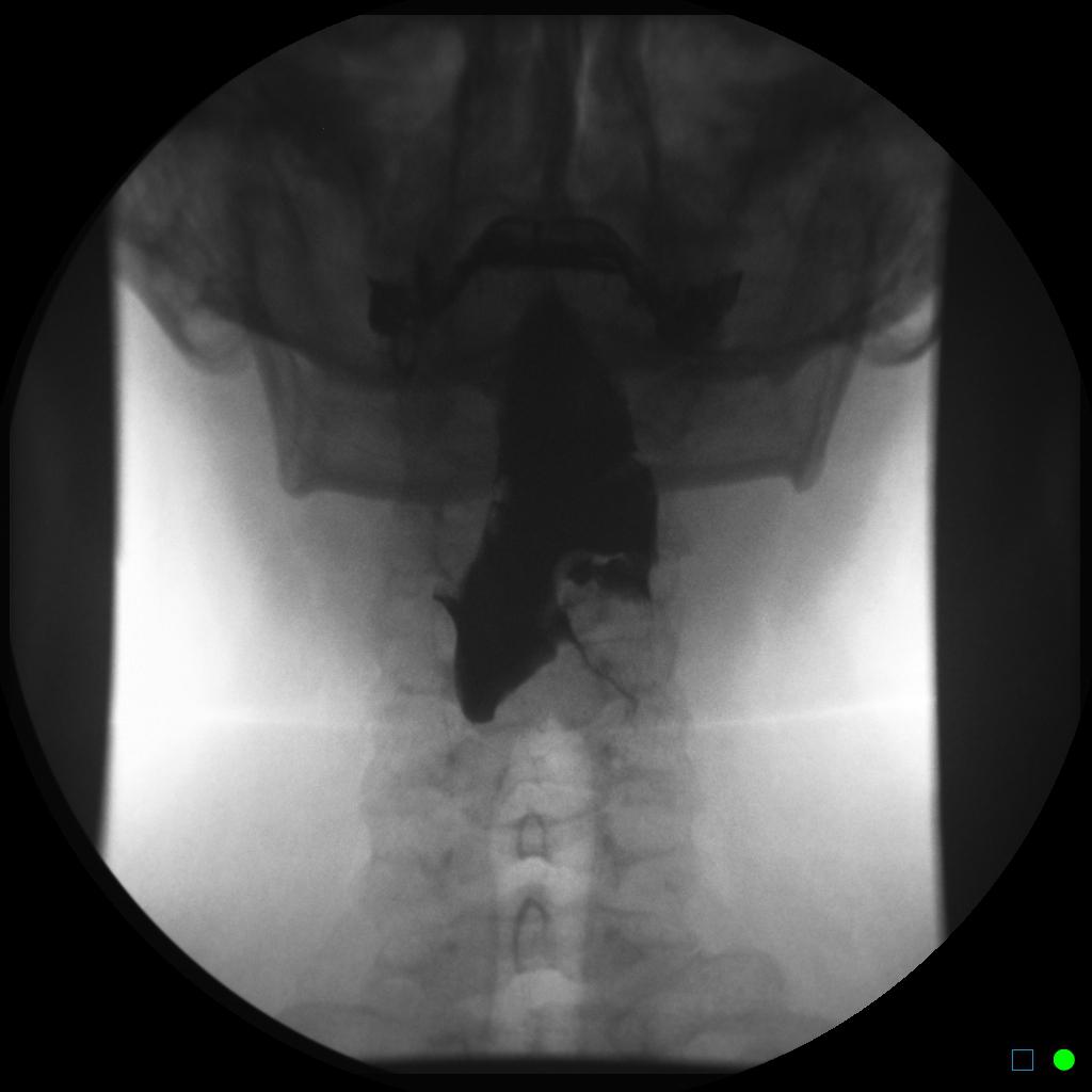 Left-piriform-fossa-mass-likely-scc (2).jpg Faizan Sheraz
16:01, 30 September 2015
1,024 × 1,024; 50 KB
-
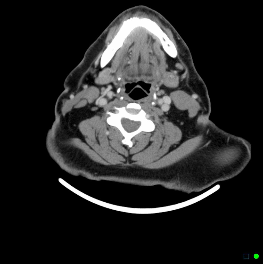
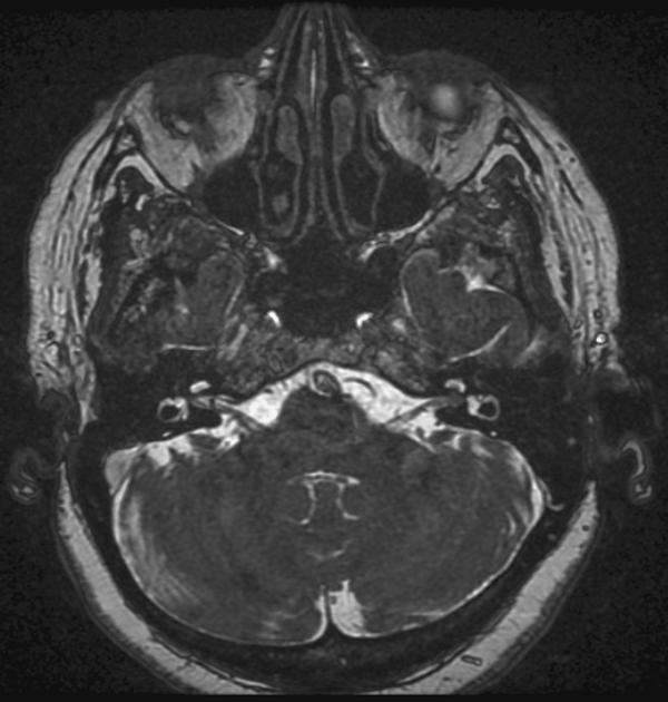
 Left-piriform-fossa-mass-likely-scc (1).jpg Faizan Sheraz
15:56, 30 September 2015
1,024 × 1,024; 50 KB
-
-
-

-
-
-
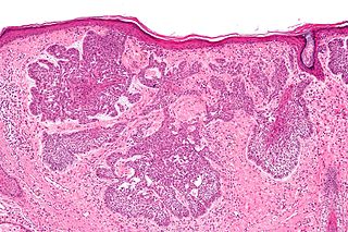
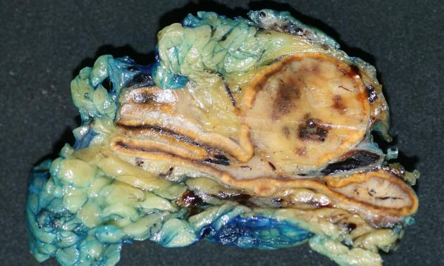
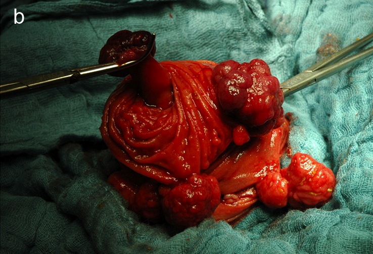
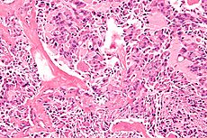
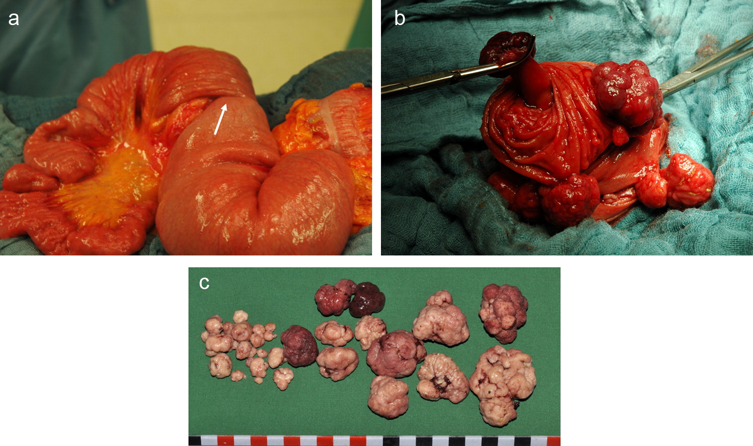
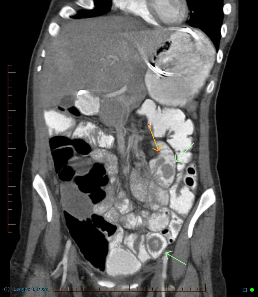
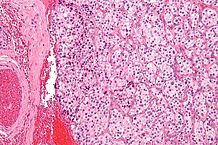
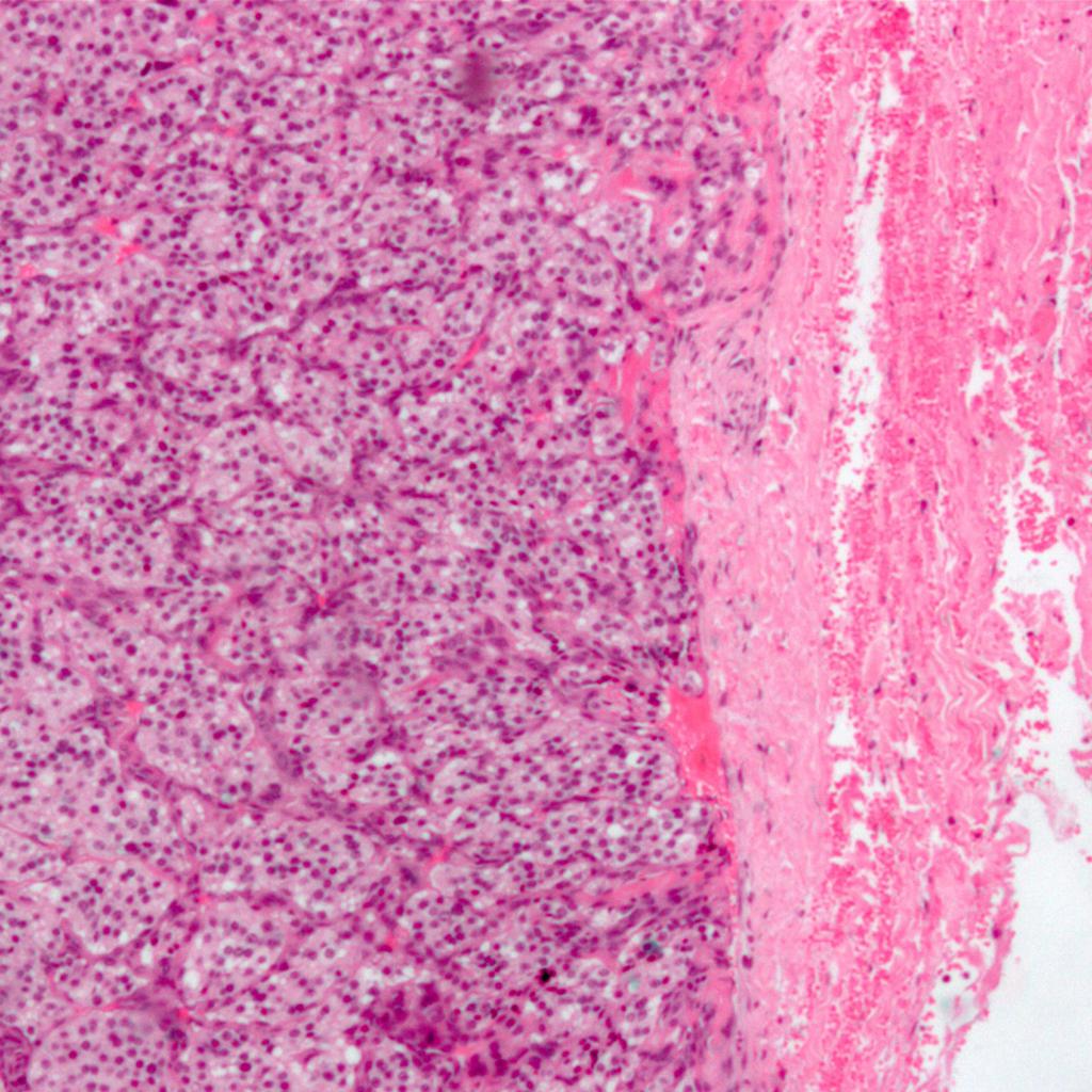
 Terminal-ileum-carcinoid-well-differentiated-neuroendocrine-tumour (1).JPG Parminder Dhingra
20:42, 29 September 2015
948 × 1,024; 116 KB
-
-
-
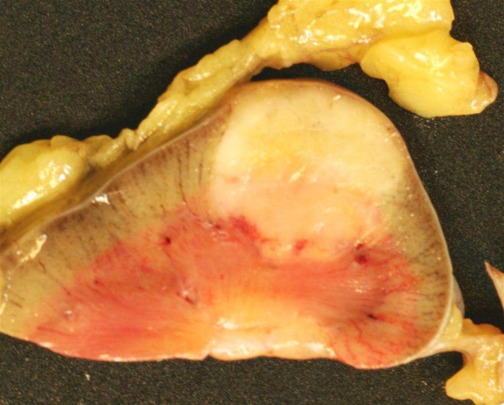
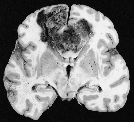
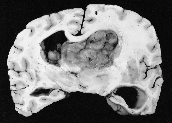
 Medulloblastoma gross pathology Dr Frank Gaillard.jpg Haytham Allaham
15:56, 29 September 2015
442 × 442; 31 KB
-
-
 Medulloblastoma Partial MAP2 immunoreactivity..jpg Haytham Allaham
11:35, 29 September 2015
120 × 89; 6 KB
-
 Medulloblastoma Epitheloid ribboning and nuclear moulding of tumor cells..jpg Haytham Allaham
11:33, 29 September 2015
120 × 89; 6 KB
-
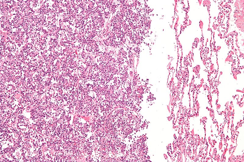
 Medulloblastoma Areas of geographic necrosis..jpg Haytham Allaham
11:32, 29 September 2015
120 × 89; 6 KB
-
-
-
-
-
-
-
-
-
-
-
-
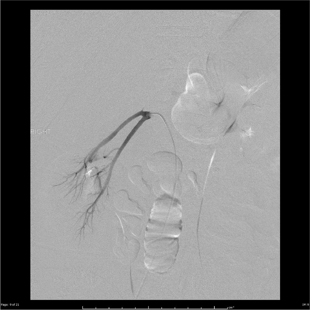
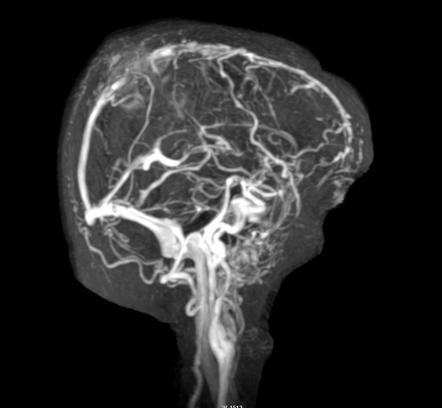 Angiography meningioma Dr Bruno Di Muzio.JPG Haytham Allaham
19:23, 28 September 2015
442 × 408; 17 KB
-
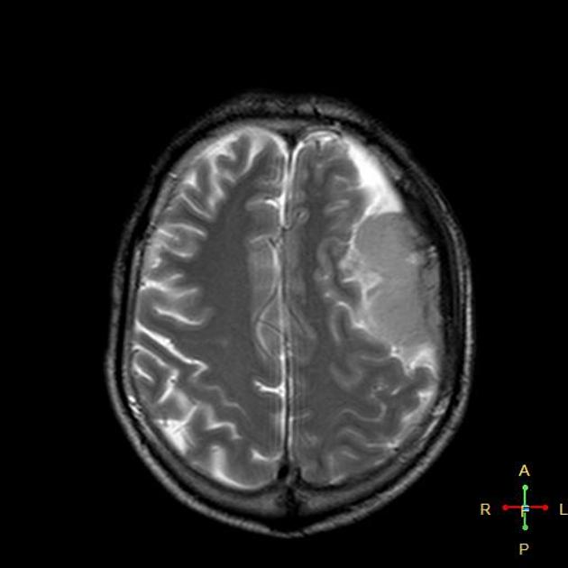
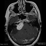
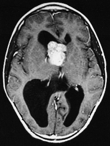
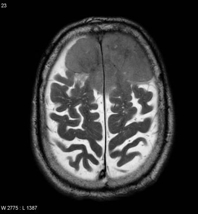 MRI meningioma anaplastic Dr Frank Gaillard.jpg Haytham Allaham
19:16, 28 September 2015
410 × 442; 20 KB
-
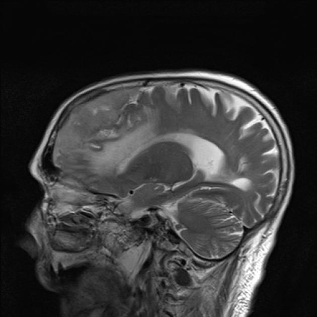
 MRI meningioma with vascular pedicle Dr Frank Gaillard.jpg Haytham Allaham
19:12, 28 September 2015
427 × 442; 21 KB
-
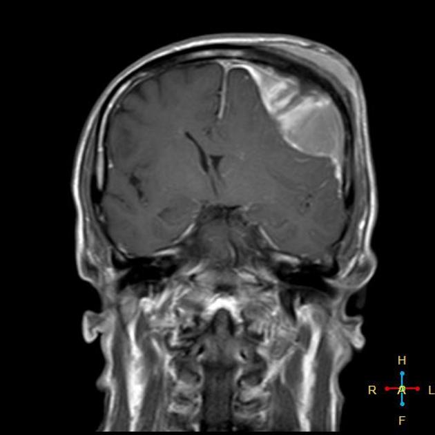
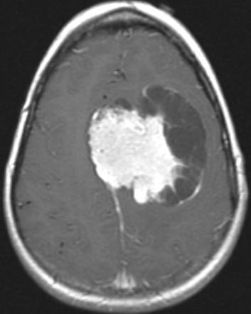
 MRI invasive meningioma Dr Bita Abbasi.jpeg Haytham Allaham
19:11, 28 September 2015
630 × 630; 36 KB
-
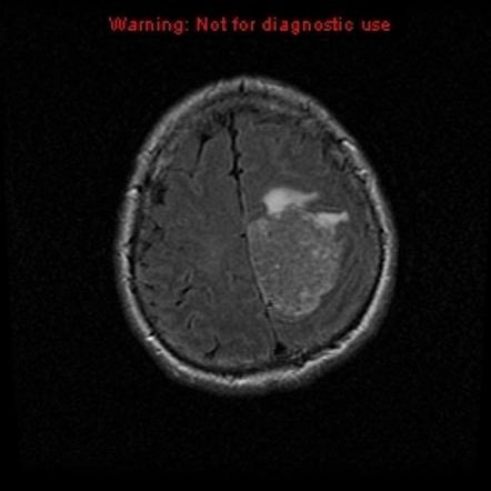
 MRI malignant meningioma Dr Ahmed Abd Rabou.jpg Haytham Allaham
19:03, 28 September 2015
630 × 630; 32 KB
-

-
 MRI meningioma dural tail sign Dr Frank Gaillard.jpg Haytham Allaham
18:59, 28 September 2015
630 × 630; 30 KB
-
 MRI meningioma cystic Dr Frank Gaillard.jpg Haytham Allaham
18:57, 28 September 2015
353 × 442; 15 KB
-
 MRI olfactory meningioma Dr Frank Gaillard.jpg Haytham Allaham
18:55, 28 September 2015
504 × 630; 42 KB
-
-
-
-

-
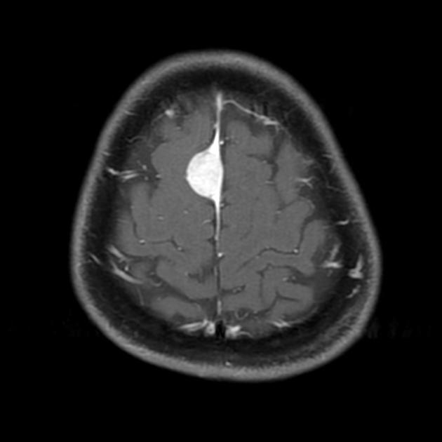
 MRI convexity meningioma Dr Sajoscha Sorrentino.jpg Haytham Allaham
13:42, 28 September 2015
511 × 630; 21 KB
-
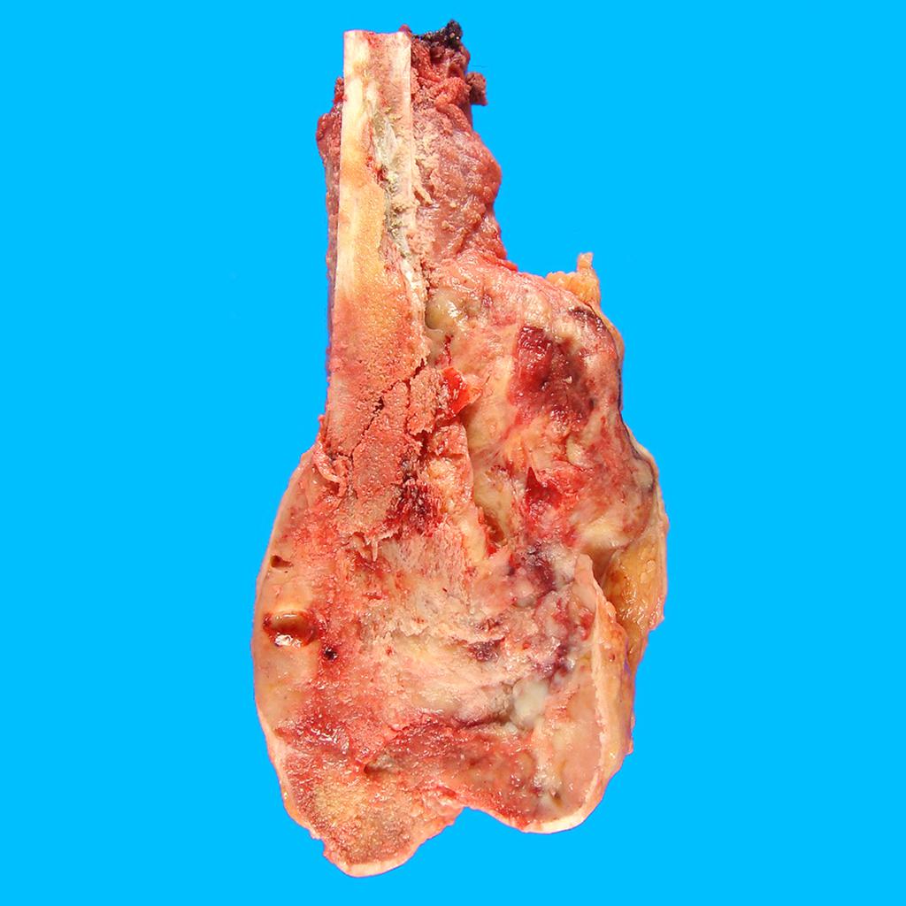
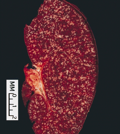
 Osteosarcoma gross pathology.jpg Suveenkrishna Pothuru
13:10, 28 September 2015
1,024 × 1,024; 94 KB
-
-

-
-
-
 CT scan of cerbral convexity meningioma.jpg Haytham Allaham
20:48, 26 September 2015
630 × 504; 31 KB
-

-
 CT scan of cerbellopontine angle meningioma.jpg Haytham Allaham
20:43, 26 September 2015
596 × 630; 35 KB
-
-

-

-

-

-

-

-
-
-

-
-
-

-
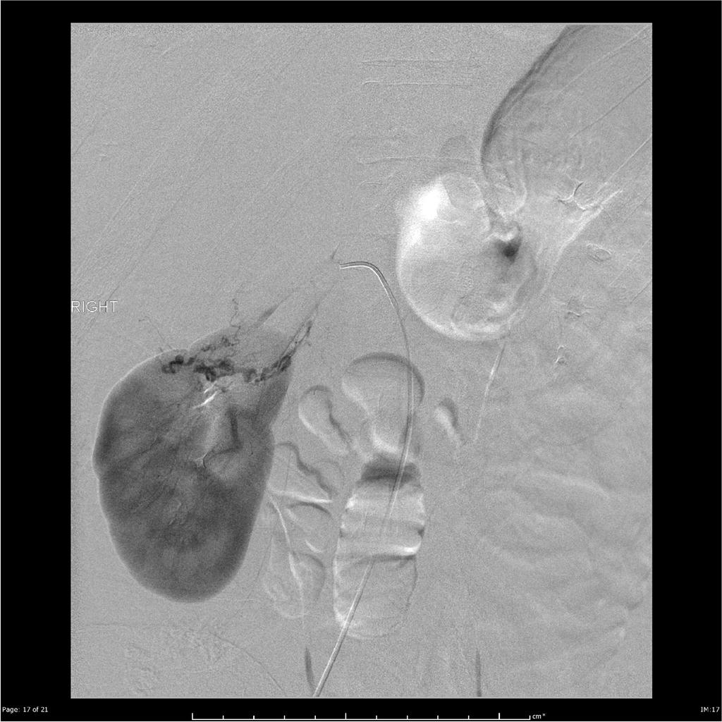
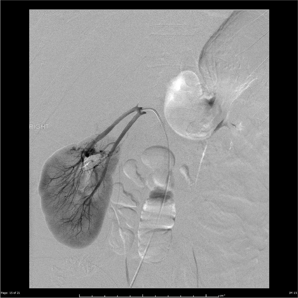
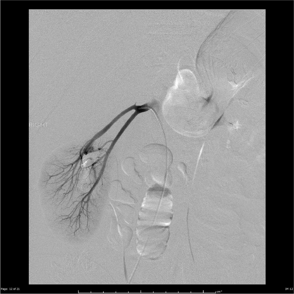
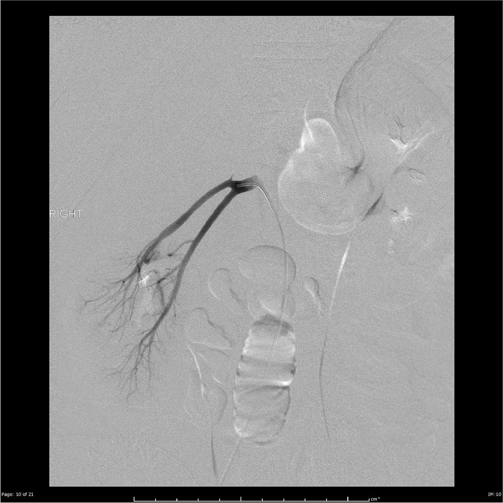
 Retroperitoneal-haemorrhage-from-renal-angiomyolipoma.jpg Faizan Sheraz
19:59, 25 September 2015
1,024 × 848; 91 KB
-
-

-
-
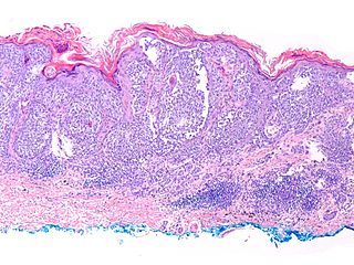
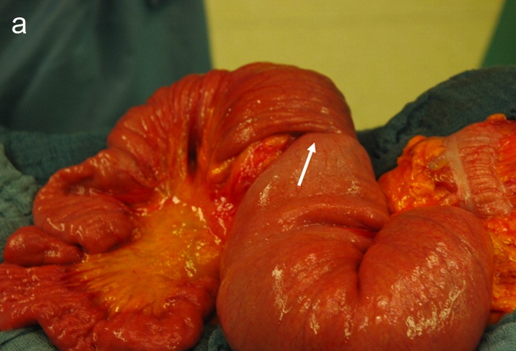
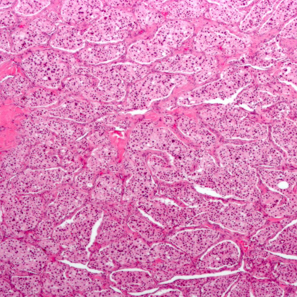
 Terminal-ileum-carcinoid-well-differentiated-neuroendocrine-tumour.jpg Parminder Dhingra
19:18, 25 September 2015
1,024 × 768; 137 KB
-
-
-
-
-
-
-

-
-

-
-
-

-
-
-
-
-

-
-
-
-
-
-
-
-
-
-
-
-
-
-
-
-
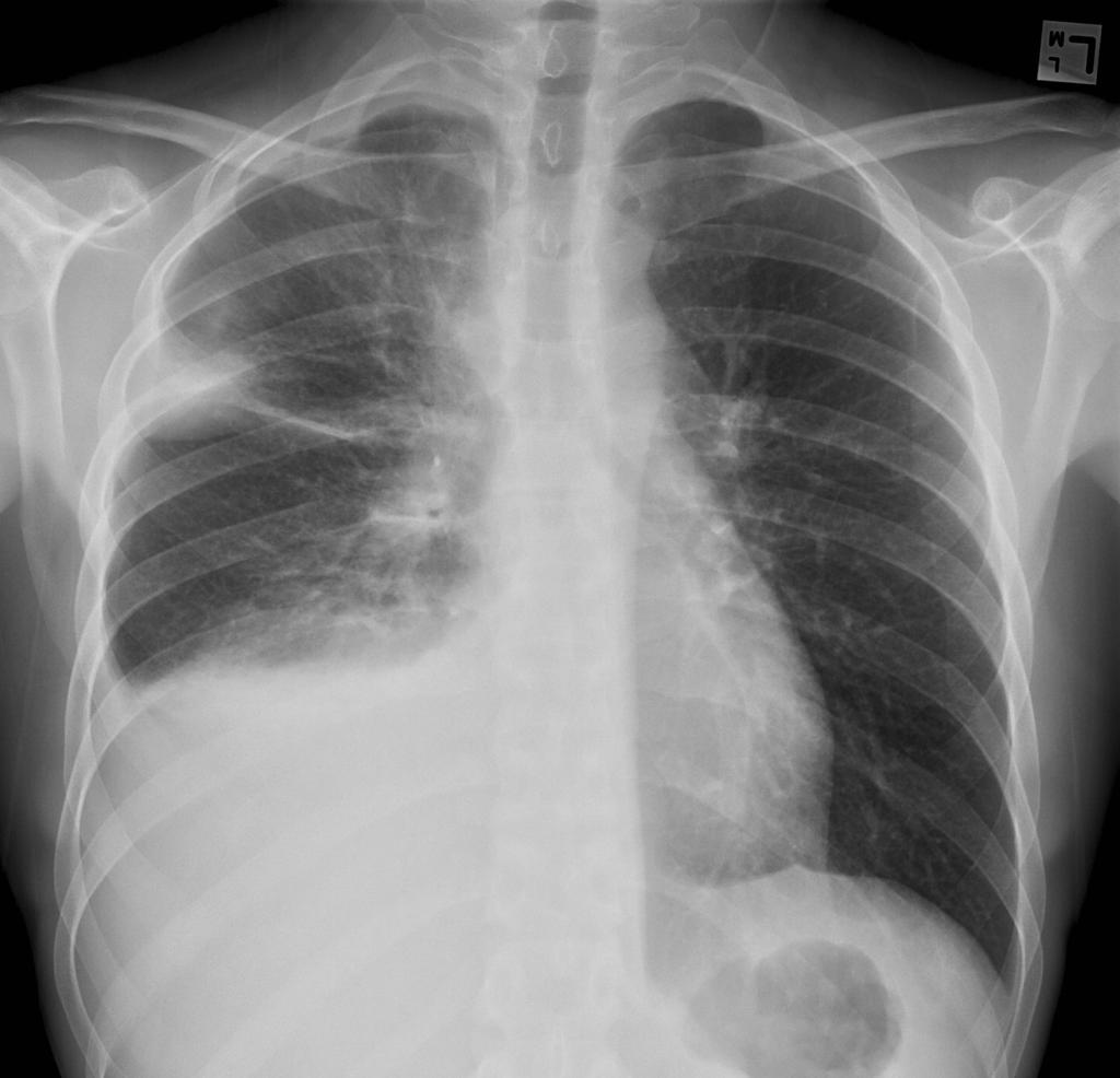
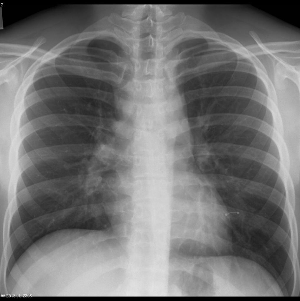
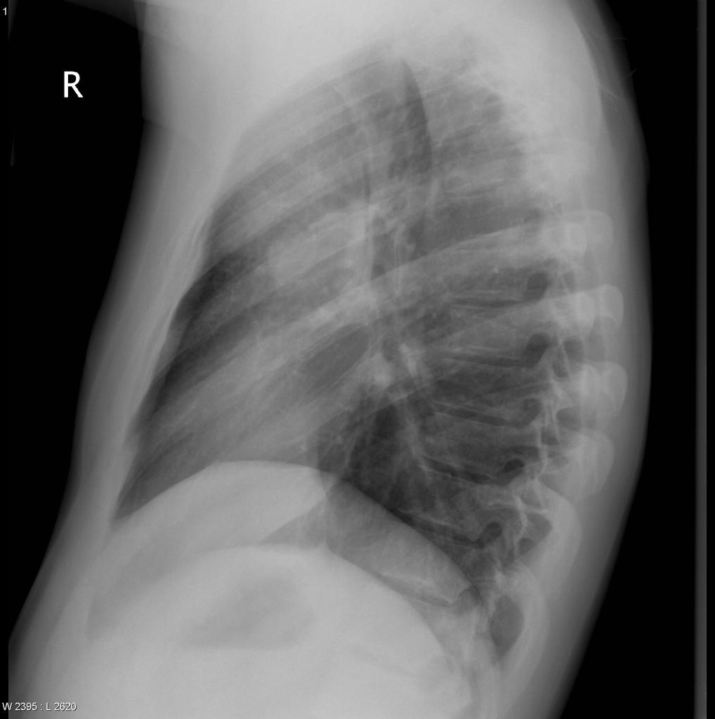 Hodgkin's lymphoma Lateral view Chest X ray .jpg Sowminya Arikapudi
18:04, 24 September 2015
1,018 × 1,024; 55 KB
-
-
-
-
-
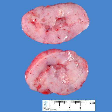
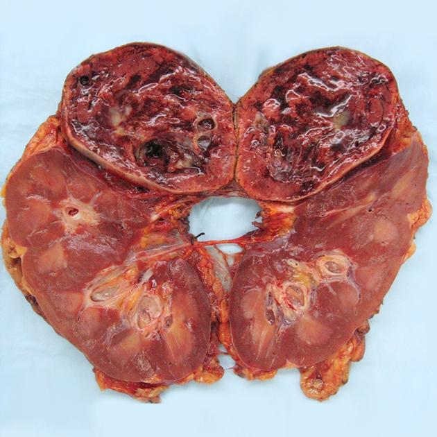
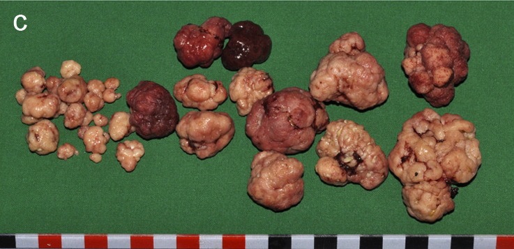
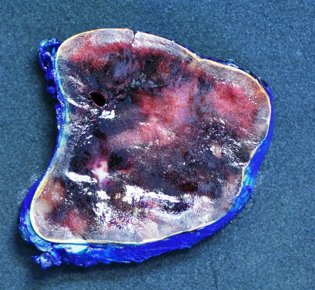
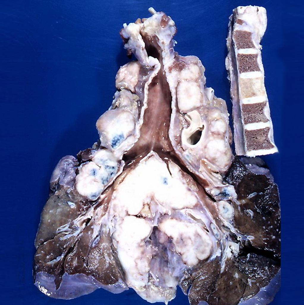 Hodgkin's lymphoma Gross Pathology.jpg Sowminya Arikapudi
15:49, 24 September 2015
1,023 × 1,024; 143 KB
-
-
-
-
-
-
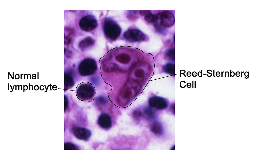
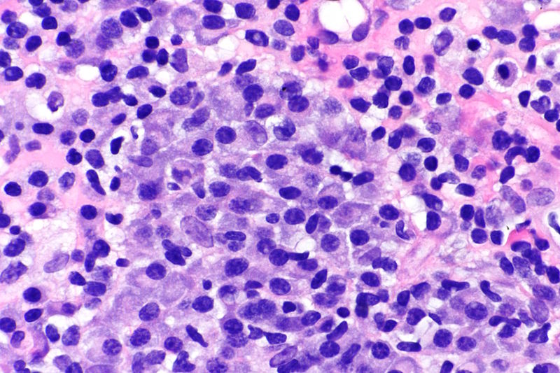
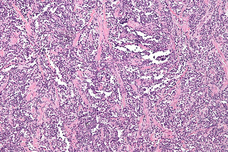
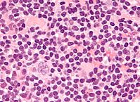
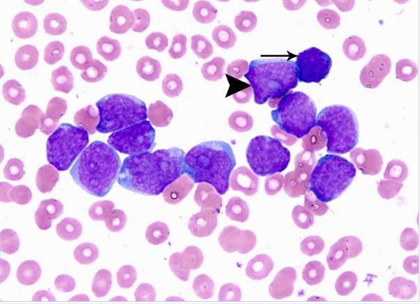
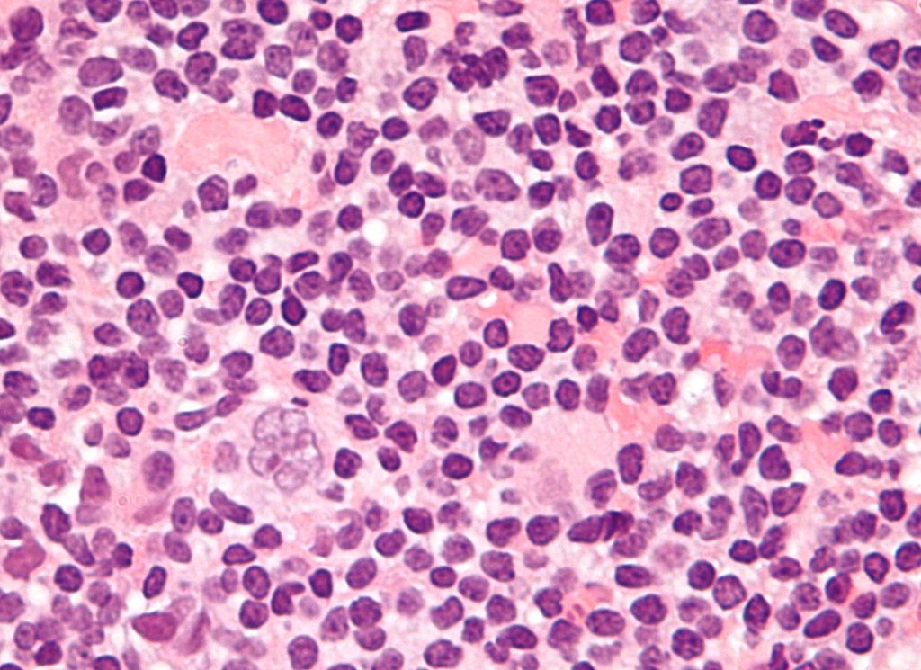 Popcorn cell in nodular lymphocyte predominant Hodgkin lymphoma.jpg Sowminya Arikapudi
20:04, 23 September 2015
1,024 × 745; 147 KB
-
-
-
 Osteosarcoma distal femur pathology.jpg Suveenkrishna Pothuru
18:43, 23 September 2015
1,024 × 1,024; 94 KB
-

-
-
-

-
 Multiple myeloma treatment algorithim.png Haytham Allaham
22:32, 22 September 2015
792 × 610; 160 KB
-
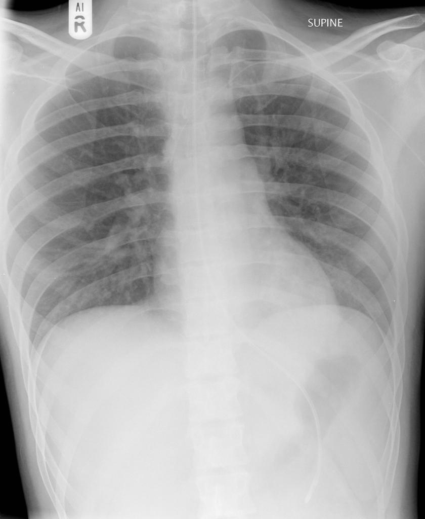
 Non-Hodgkin lymphoma chest-x ray.jpeg Sowminya Arikapudi
20:35, 22 September 2015
1,024 × 802; 159 KB
-
-
-
-
-
-

-
-
-
-
-
-
-
-

-
-
-

-
-
-
-
-
-

-
-
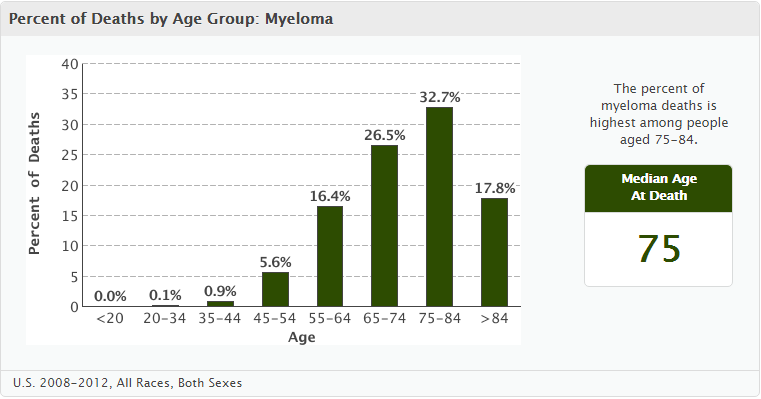 Percent od deaths per age group Myeloma.png Haytham Allaham
15:18, 21 September 2015
761 × 399; 40 KB
-
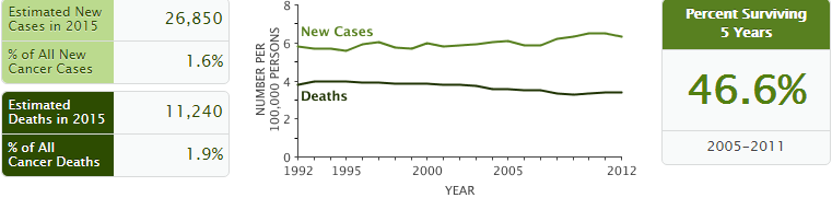
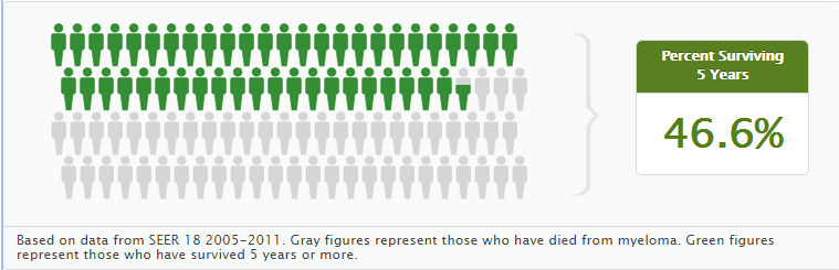
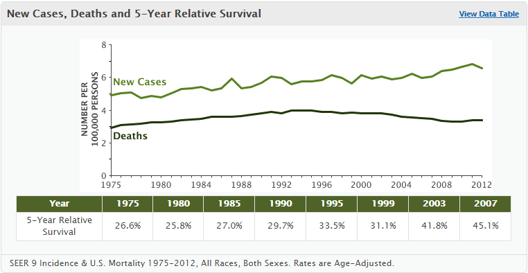 New Cases, Deaths and 5-Year Relative Survival.png Haytham Allaham
15:08, 21 September 2015
762 × 394; 38 KB
-
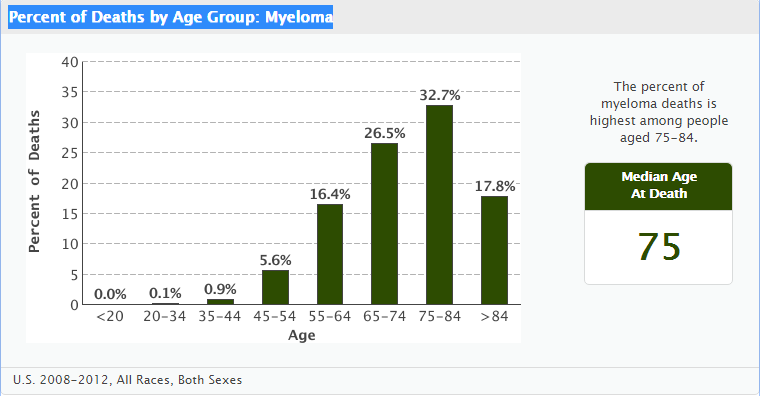 Percent of Deaths by Age Group Myeloma.png Haytham Allaham
15:08, 21 September 2015
760 × 396; 40 KB
-
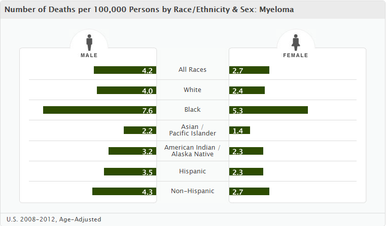 Number of Deaths per 100,000 Persons by RaceEthnicity & Sex Myeloma.png Haytham Allaham
15:08, 21 September 2015
761 × 447; 42 KB
-
-
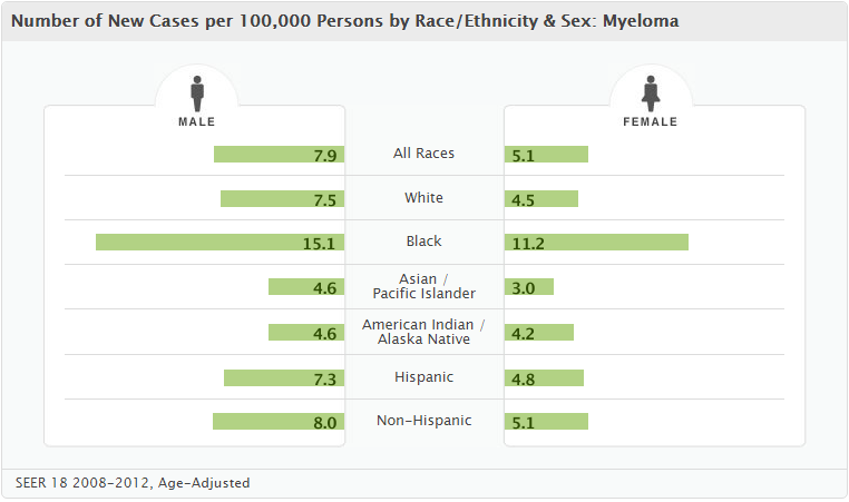 Number of new cases per race and gender.png Haytham Allaham
14:41, 21 September 2015
762 × 451; 42 KB
-

-
-

-
-

-
 Plexiform schwannoma with high magnifaction.jpg Simrat Sarai
02:39, 21 September 2015
120 × 98; 4 KB
-
-
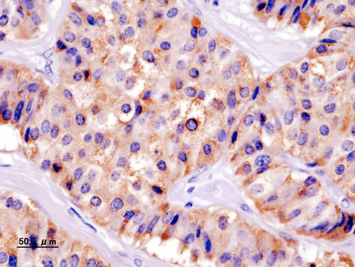
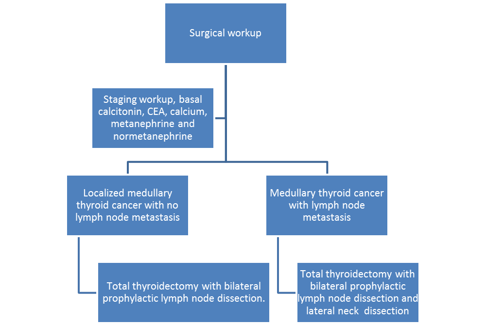
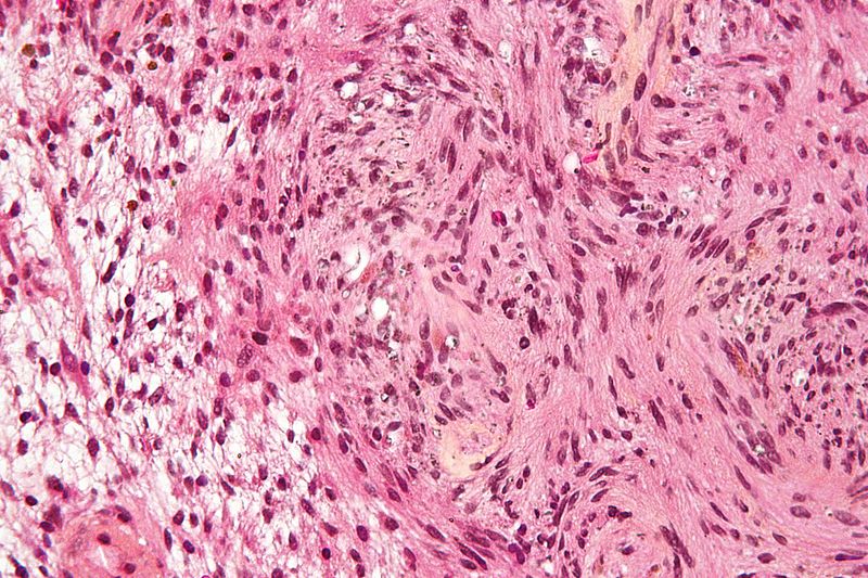
 MIcroscopic pathology of medullary thyroid cancer .jpg Ammu Susheela
19:51, 20 September 2015
230 × 153; 20 KB
-

-

-
-
-
-
-
-
-

-

-

-

-

-

-
-
-
-
-
-

-
-
-
-

-

-
 5907de80-c98d-4eba-8273-7a771d9ee9b5.jpg Sujit Routray
15:15, 12 September 2015
1,080 × 1,069; 107 KB
-

-

-

-

-
-
-
-
-
-

-

-
 97px-Hodgkin's Disease (Essentials of Medicine).jpg Sowminya Arikapudi
19:22, 8 September 2015
97 × 145; 4 KB
-

-

-
-
-

-
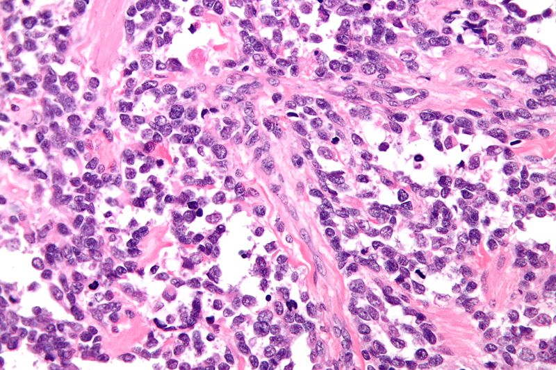
 Alveolar rhabdomyosarcoma - intermed mag.jpg Suveenkrishna Pothuru
16:56, 8 September 2015
800 × 533; 226 KB
-
 800px-Alveolar rhabdomyosarcoma - intermed mag.jpg Suveenkrishna Pothuru
16:55, 8 September 2015
800 × 533; 226 KB
-
-
 199px-Popcorn cell in nodular lymphocyte predominant Hodgkin lymphoma - very high mag cropped.jpg Sowminya Arikapudi
19:33, 4 September 2015
199 × 145; 15 KB
-
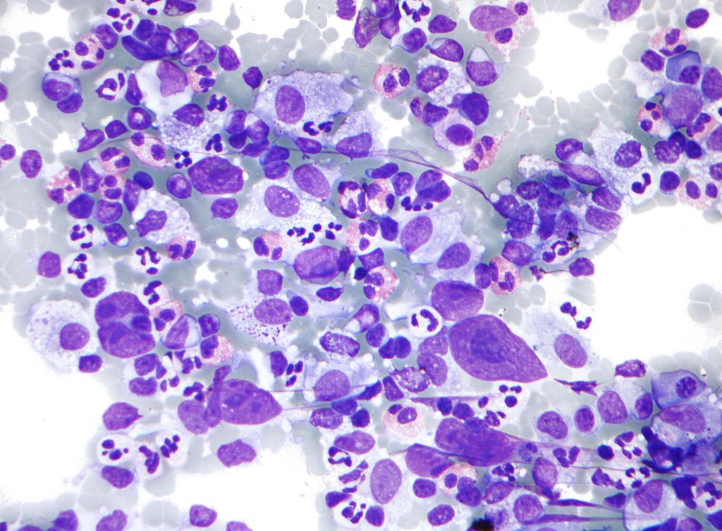
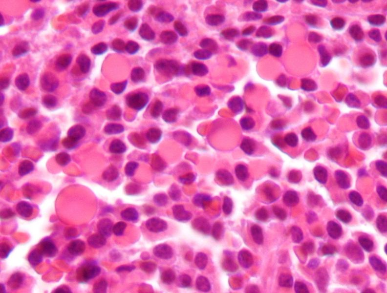
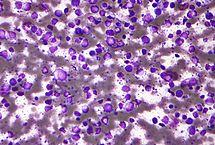
 197px-Hodgkin lymphoma cytology large.jpg Sowminya Arikapudi
19:31, 4 September 2015
197 × 145; 14 KB
-
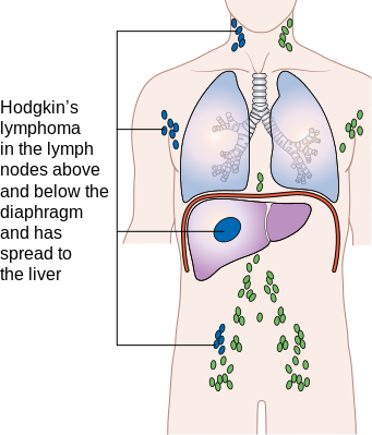
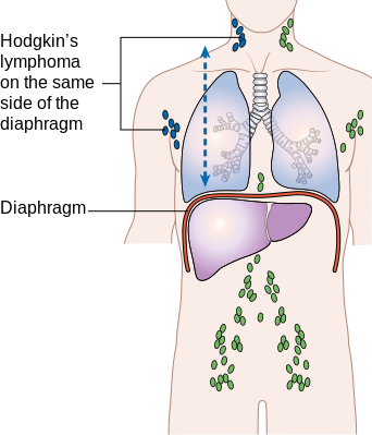
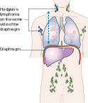
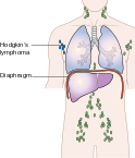
 124px-Diagram showing stage 4 Hodgkin's lymphoma CRUK 230.svg.png Sowminya Arikapudi
18:34, 4 September 2015
124 × 145; 14 KB
-
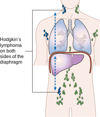
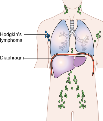
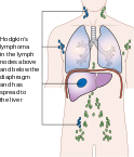
 124px-Diagram showing stage 3 Hodgkin's lymphoma CRUK 221.svg.png Sowminya Arikapudi
18:33, 4 September 2015
124 × 145; 14 KB
-
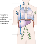
 124px-Diagram showing stage 2 Hodgkin's lymphoma CRUK 208.svg.png Sowminya Arikapudi
18:31, 4 September 2015
124 × 145; 14 KB
-
 124px-Diagram showing stage 1 Hogkin's lymphoma CRUK 191.svg.png Sowminya Arikapudi
18:29, 4 September 2015
124 × 145; 13 KB
-
-
-

-
-

-
-

-

-
 800px-Alveolar rhabdomyosarcoma - very high mag.jpg Suveenkrishna Pothuru
15:14, 1 September 2015
800 × 533; 129 KB
-
-

-
 Normal-nasogastric-tube-position-annotated.jpg William J Gibson
18:38, 31 August 2015
840 × 1,024; 79 KB
-
-
 Endometrioid endometrial adenocarcinoma very high mag.jpg Monalisa Dmello
18:10, 31 August 2015
640 × 427; 96 KB
-
 320px-Endometrioid endometrial adenocarcinoma very high mag2.jpg Monalisa Dmello
18:08, 31 August 2015
320 × 213; 31 KB
-
 218px-Mantle cell lymphoma - low mag - cyclin D1.jpg Sowminya Arikapudi
15:44, 31 August 2015
218 × 145; 10 KB
-
-
 193px-Nodular Mantle Cell Lymphoma - low power view - by Gabriel Caponetti.jpg Sowminya Arikapudi
15:31, 31 August 2015
193 × 145; 6 KB
-
 193px-Nodular Mantle Cell Lymphoma - high power view - by Gabriel Caponetti.jpg Sowminya Arikapudi
15:22, 31 August 2015
193 × 145; 14 KB
-
 97px-Mantle cell lymphoma - intermed mag.jpg Sowminya Arikapudi
15:18, 31 August 2015
97 × 146; 6 KB
-
-
-
 218px-Carotid body tumour 2 intermed mag.jpg Ahmad Al Maradni
13:16, 31 August 2015
218 × 145; 17 KB
-
-
-

-

-

-

-

-
-
 800px-Bone Chondrosarcoma Dedifferentiated HP PA.jpg Suveenkrishna Pothuru
15:09, 28 August 2015
800 × 600; 118 KB
-
 707px-Bone Chondrosarcoma Dedifferentiated PA copy.jpg Suveenkrishna Pothuru
15:07, 28 August 2015
707 × 599; 187 KB
-
-

-

-
-

-
 800px-Bone Chondrosarcoma Mesenchymal MP5 PA.JPG Suveenkrishna Pothuru
21:07, 27 August 2015
800 × 597; 173 KB
-
-
 800px-Bone Chondrosarcoma ClearCell MP PA.jpg Suveenkrishna Pothuru
20:55, 27 August 2015
800 × 600; 157 KB
-

-
 800px-Bone Chondrosarcoma ClearCell HP PA.jpg Suveenkrishna Pothuru
17:57, 27 August 2015
800 × 600; 162 KB
-
 800px-Bone Chondrosarcoma Myxoid MP PA.JPG Suveenkrishna Pothuru
17:51, 27 August 2015
800 × 600; 155 KB
-
 790px-Bone Chondrosarcoma Myxoid MP2 PA.JPG Suveenkrishna Pothuru
17:43, 27 August 2015
790 × 599; 130 KB
-
 800px-Bone Chondrosarcoma Grade3 HP PA.JPG Suveenkrishna Pothuru
17:31, 27 August 2015
800 × 600; 151 KB
-
 800px-Bone Chondrosarcoma Grade2 HP PA.jpg Suveenkrishna Pothuru
17:26, 27 August 2015
800 × 600; 134 KB
-
 800px-Bone Chondrosarcoma Grade1 HP2 PA.JPG Suveenkrishna Pothuru
17:21, 27 August 2015
800 × 600; 63 KB
-

-
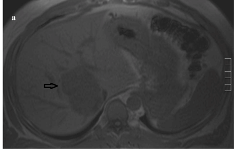
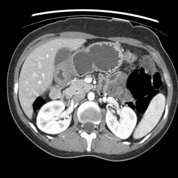
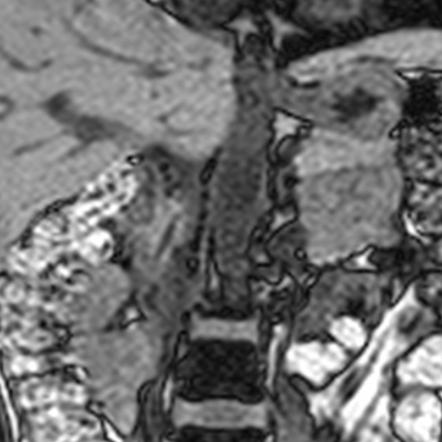
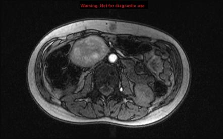
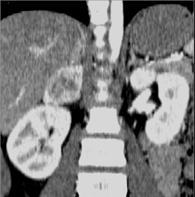
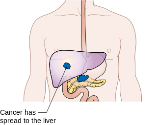
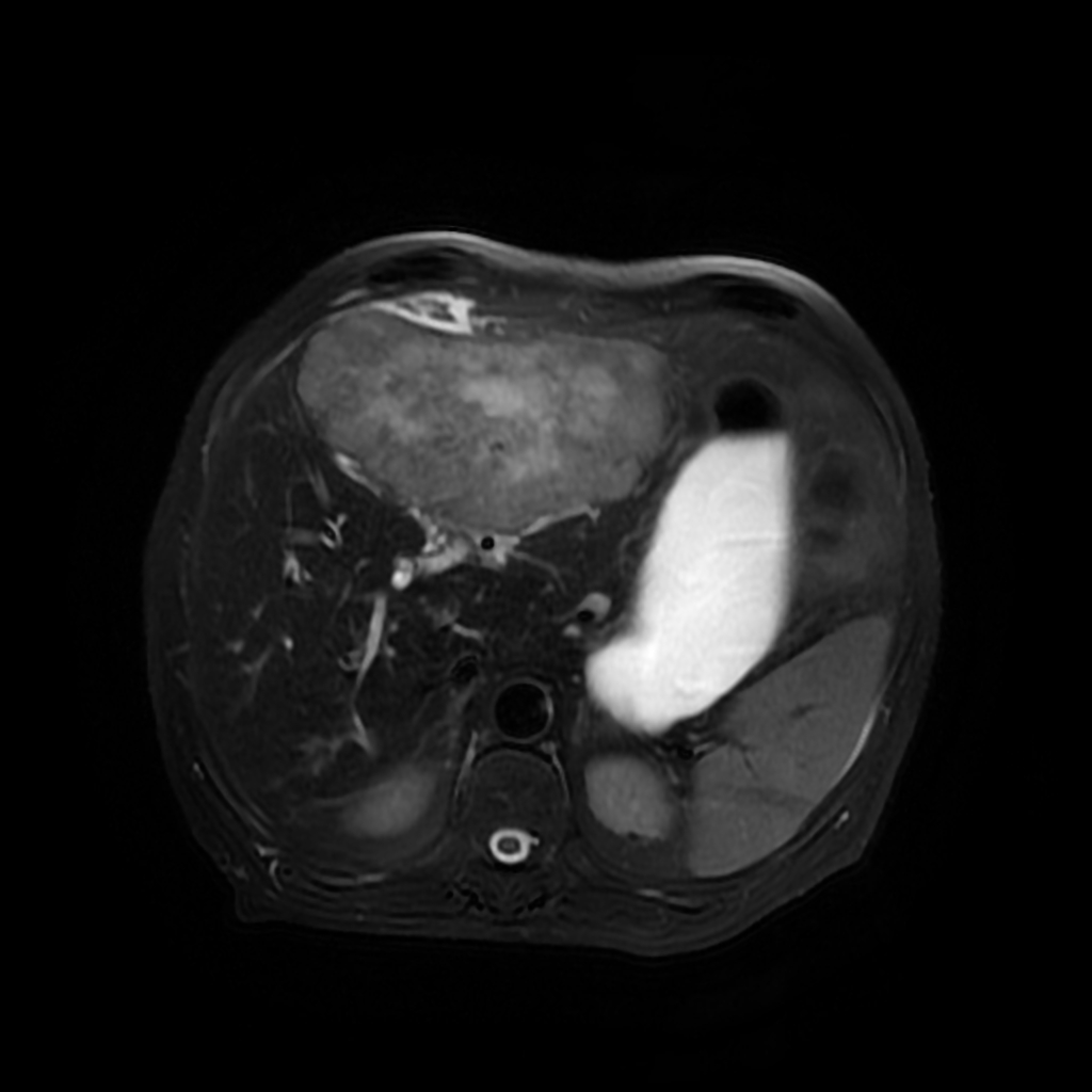 Hepatic-metastases-from-colon-carcinoma-triphasic-mri(1).jpg Mohamad Alkateb
14:51, 26 August 2015
1,024 × 1,024; 156 KB
-
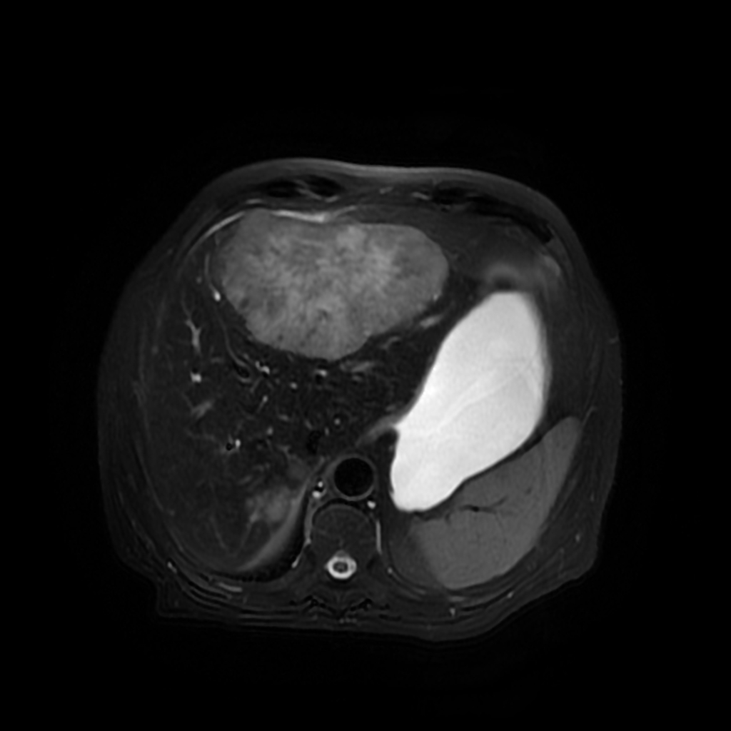 Hepatic-metastases-from-colon-carcinoma-triphasic-mri.jpg Mohamad Alkateb
14:40, 26 August 2015
1,024 × 1,024; 150 KB
-
 215px-Diffuse large B cell lymphoma - cytology low mag.jpg Sowminya Arikapudi
21:43, 25 August 2015
215 × 145; 16 KB
-
-
-
-
-
-

-

-
 Cervical Cancer Screening Guidelines for Average-Risk Women.jpg Monalisa Dmello
21:12, 21 August 2015
1,393 × 1,389; 619 KB
-
-
-
-
-

-
-
-
-
-
-
-
-
-

-
 640px-Malignant peripheral nerve sheath tumour - high mag.jpg Suveenkrishna Pothuru
23:45, 20 August 2015
640 × 427; 138 KB
-
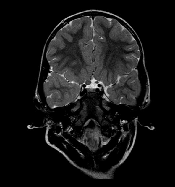
 Ependymoma of 4th ventricle in MRI1.jpg Suveenkrishna Pothuru
21:22, 20 August 2015
838 × 998; 82 KB
-

-

-

-

-

-

-
-
-
-
-
-
-

-
-

-

-

-

-

-
-

-
-
-
-
-
-
-
-

-

-

-
-
-

-
-
-
-
-
-

-
-

-
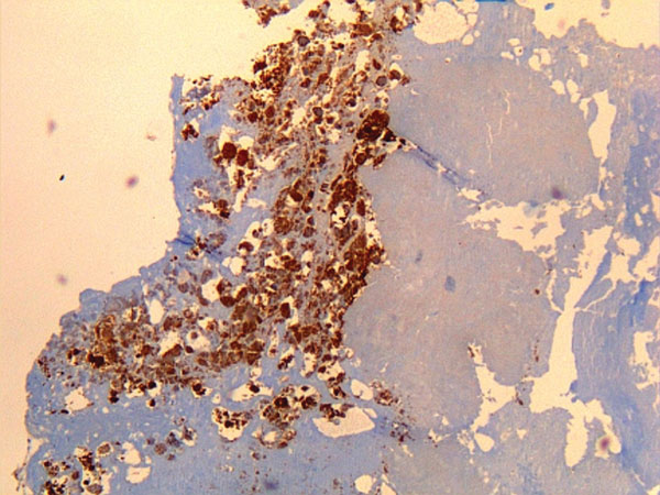 Immunohistochemical detection of Coxiella burnetii in resected cardiac valve of a 60-year-old man with Q fever endocarditis.jpg Monalisa Dmello
20:09, 3 August 2015
600 × 450; 85 KB
-
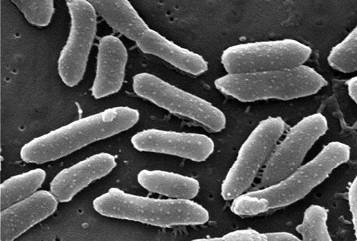
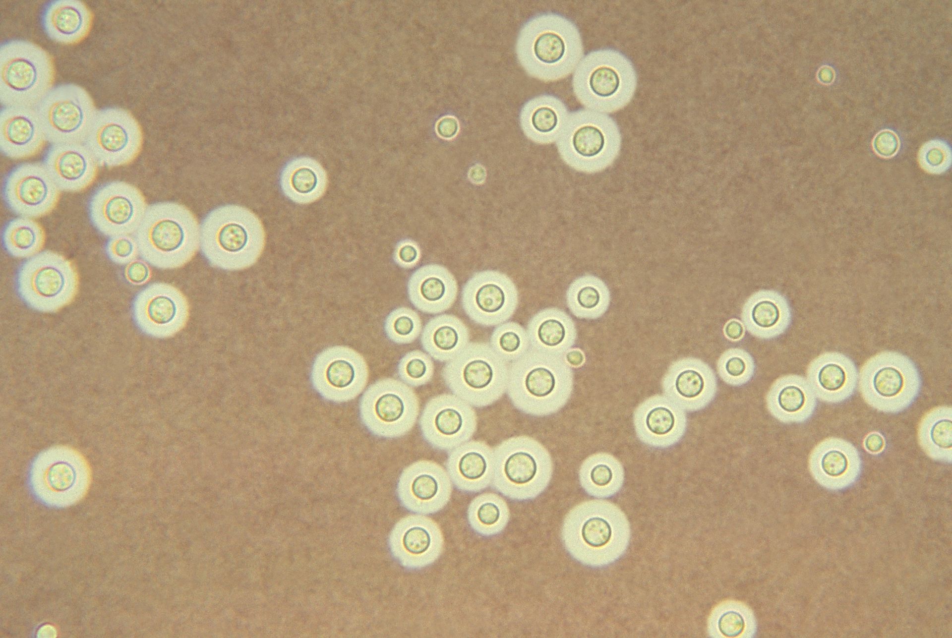
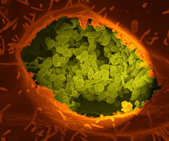 Coxiella burnetii, the bacteria that causes Q Fever.jpg Monalisa Dmello
19:51, 3 August 2015
573 × 480; 78 KB
-

-
-

-
-
-
-
-
-
-

-

-
-
-
-
-
-
-
-

-

-

-
-
-
-
-
-
-
-

-
-
-
-

-

-

-

-

-
-
-

-

-

-
-
-
-
-

-
-
-
-
-
-
-
-
-
-

-
-
-
-
-
-
-
-
-
-

-
-

-
-
-
-
-
-
-
-
-
-
-
-
-
-
-

-

-

-
-

-
-
-
-
-
-
-
-
-
-
-
-
-
-
-
-

-

-

-


_GH_production.jpg)
_GH_production.jpg)
.jpg)
.jpg)
.jpg)
.jpg)
.jpg)


.jpg)


.jpg)
.jpg)
.jpg)
.jpg)


.JPG)

.jpg)


.jpg)


.jpg)
.jpg)
.jpg)
.jpg)
.jpg)


.jpg)


.jpg)

.jpeg)
.jpg)


.gif)

.jpg)


.jpg)
.jpg)


.jpg)
.jpg)


_transitional_type.jpg)