Gallery of new files
Jump to navigation
Jump to search
This special page shows the last uploaded files.
-

-

-
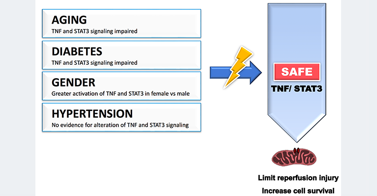
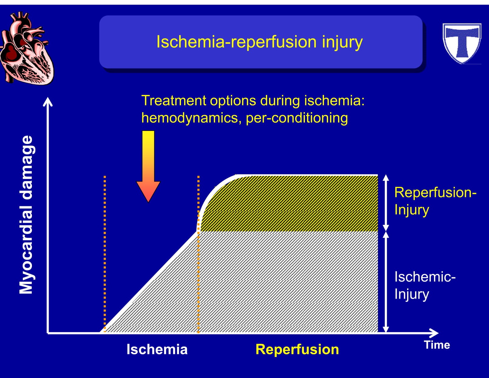
 Reperfusion injury management - Via pre-conditioning & Post-conditioning.jpg Shivam Singla
17:17, 22 August 2020
1,800 × 1,349; 216 KB
-

-
-

-

-
-
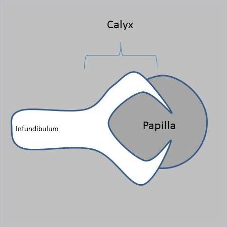
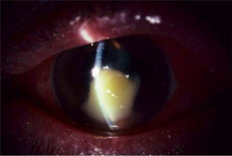
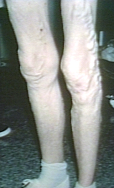
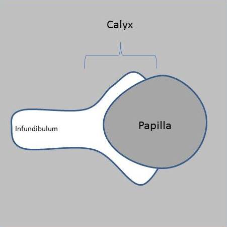 Lobster-claw-sign-papillary-necrosis-normal.jpg NNikravangolsefid
06:02, 21 August 2020
451 × 450; 11 KB
-

-
-

-

-

-

-

-

-
-
-
-
-
-
 Xray Forearm AP Lateral Madelung Deformity.jpg Akash Daswaney
13:33, 18 August 2020
1,024 × 613; 36 KB
-
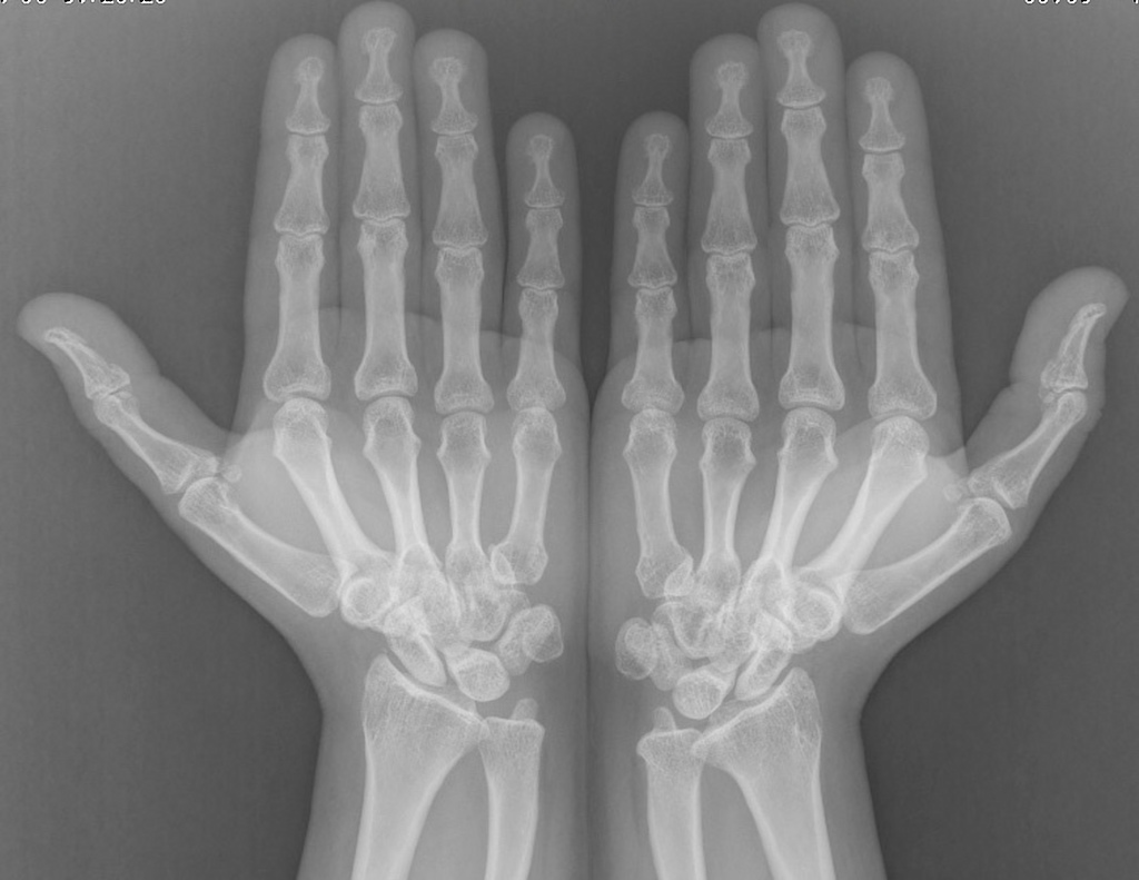
 Xray Bilateral AP view Madelung Deformity Shorted 4th Metacarpal Image 4.jpg Akash Daswaney
13:32, 18 August 2020
1,022 × 849; 46 KB
-

-
-
-
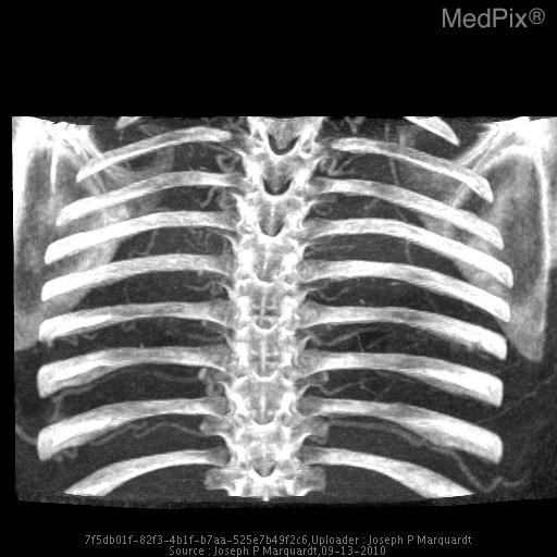 CT Angiography Enlarged Inrercoastal Arteries.jpg Akash Daswaney
13:18, 18 August 2020
512 × 512; 40 KB
-
 CT angiography Coarctation of Aorta Enlarged Internal Mammary Arteries.jpg Akash Daswaney
13:13, 18 August 2020
512 × 512; 30 KB
-
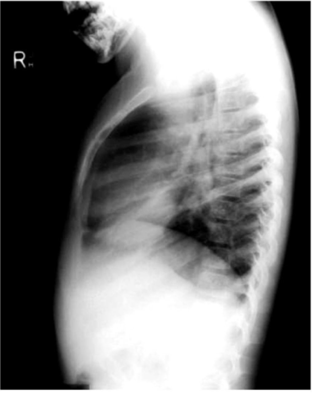
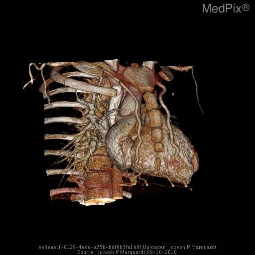
 CT Angiography Coarctation of Aorta Left Lateral Volume rendered image.jpg Akash Daswaney
12:46, 18 August 2020
512 × 512; 30 KB
-
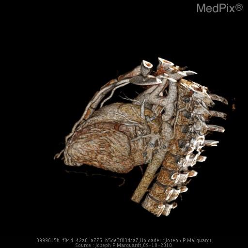
 CT Angiography Coarctation of Aorta Original Image 6.jpg Akash Daswaney
12:07, 18 August 2020
1,024 × 1,024; 96 KB
-
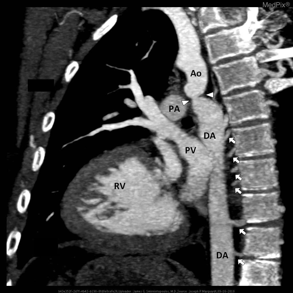 CT Angiography Coarctation of Aorta Flipped and Labelled Image 5.jpg Akash Daswaney
12:01, 18 August 2020
1,024 × 1,024; 100 KB
-
-
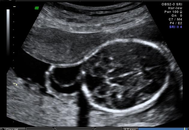
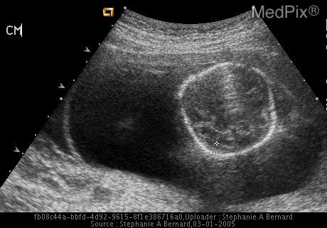 Ultrasound Cystic Hygroma with hydrops fetalis Turner Syndrome.jpg Akash Daswaney
11:38, 18 August 2020
460 × 321; 32 KB
-
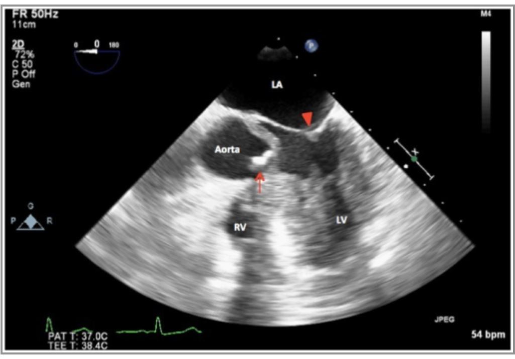 Echo Aortic Stenosis seen in Turner Syndrome.JPG Akash Daswaney
11:30, 18 August 2020
1,025 × 715; 68 KB
-

-

-

-
-
-

-

-

-
-

-

-

-

-
-

-
 Myocardial ischemia reperfusion injury.jpg Shivam Singla
17:21, 14 August 2020
1,700 × 2,200; 338 KB
-

-

-

-
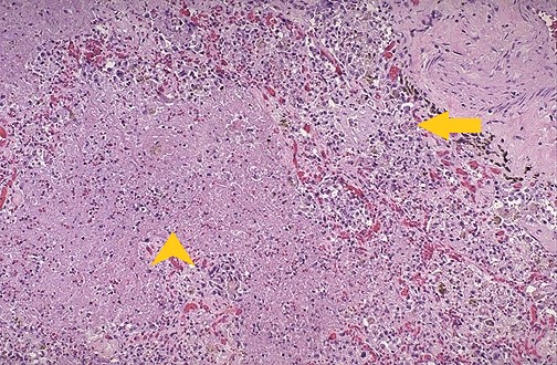
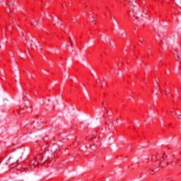
 Neutrophils involved in tissue destruction.jpg Shivam Singla
23:49, 11 August 2020
800 × 387; 217 KB
-
-

-

-

-

-

-

-
 Blausen 0057 ArtificialHeartValve StFrancis.png Javaria Anwer
20:54, 7 August 2020
472 × 790; 339 KB
-

-

-

-

-

-

-
-

-

-

-

-
-
-
-

-
-

-

-
-

-
-
-

-

-

-

-
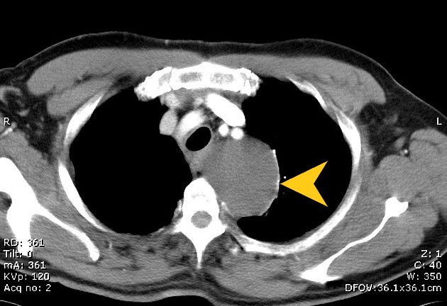
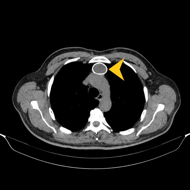
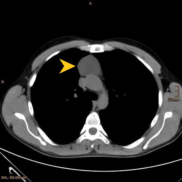
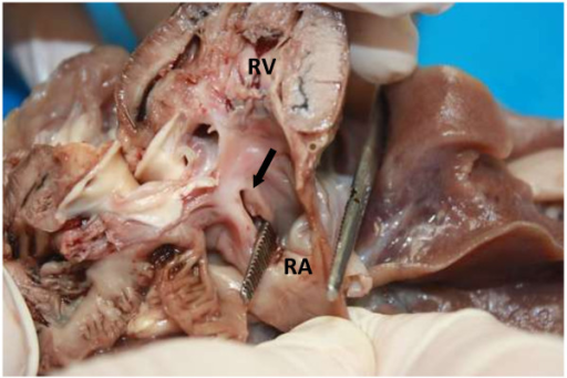
 Dilatation of pulmonary artery-image reconstrust.jpg Javaria Anwer
16:54, 4 August 2020
367 × 368; 47 KB
-
-
 Summary- Treatment for Hypernatremia based on Volume status.png MLakhmalla
14:05, 4 August 2020
755 × 615; 92 KB
-
 Right-ventricular-enlargement-on-chest-radiography-illustration lateral view.png Javaria Anwer
13:50, 4 August 2020
898 × 898; 105 KB
-
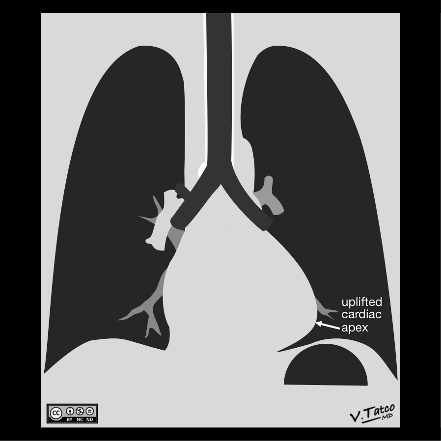 Right-ventricular-enlargement-on-chest-radiography-illustration.png Javaria Anwer
13:35, 4 August 2020
898 × 898; 79 KB
-
-

-
 Worldwide causes of acute liver failure.jpeg Roghayeh Marandi
18:07, 2 August 2020
1,536 × 1,305; 349 KB
-

-
-
-

-

-

-

-

-
-
-

-

-

-

-

-

-

-

-

-

-

-
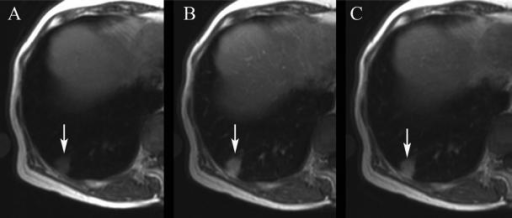
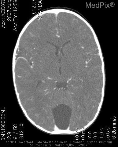
 Child with impacted metal strip (white arrow)..png Joanna Ekabua
01:40, 27 July 2020
512 × 341; 349 KB
-

-

-

-
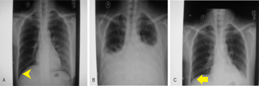
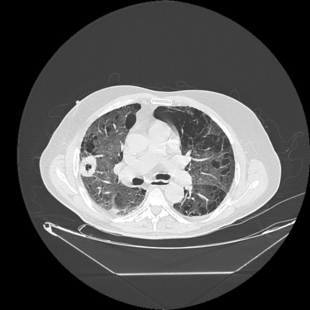
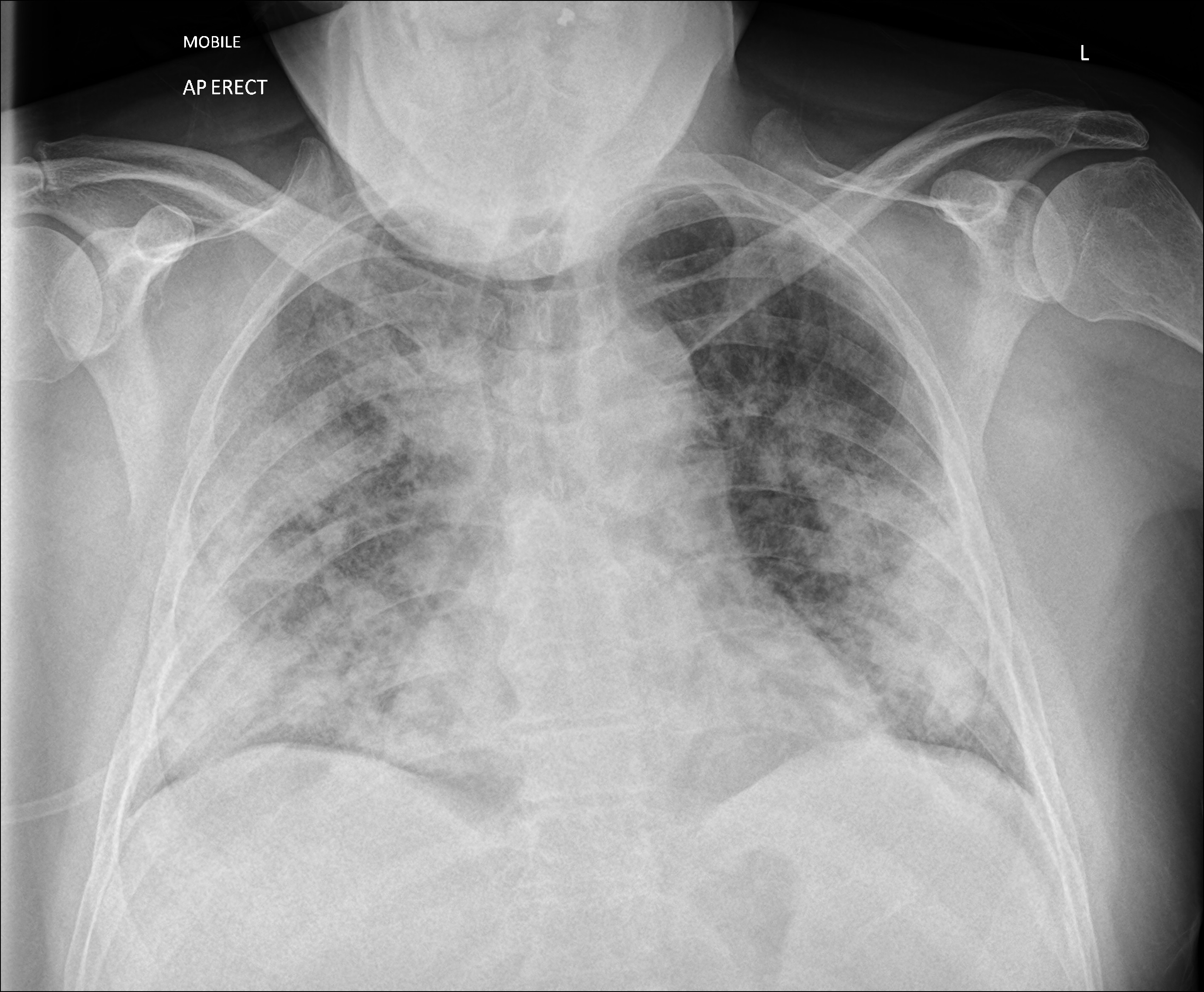
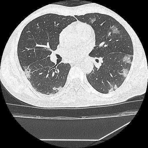
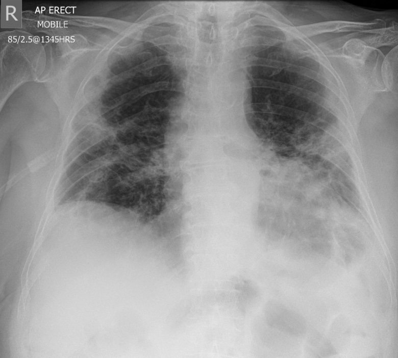
 Covid-19-rapidly-progressive-acute-respiratory-distress-syndrome-ards.jpg Ramyar
21:35, 24 July 2020
894 × 762; 78 KB
-

-
-
-
-

-
-
-
-
-

-
-
-

-
-

-
-
-

-

-
-

-

-

-

-

-

-
-

-
-
-
-

-
-

-

-

-
-

-

-

-
-

-
-

-

-
-

-

-

-

-

-

-

-

-
-
-

-

-

-
-

-

-

-
-

-

-

-

-

-

-

-

-

-

-

-

-

-

-
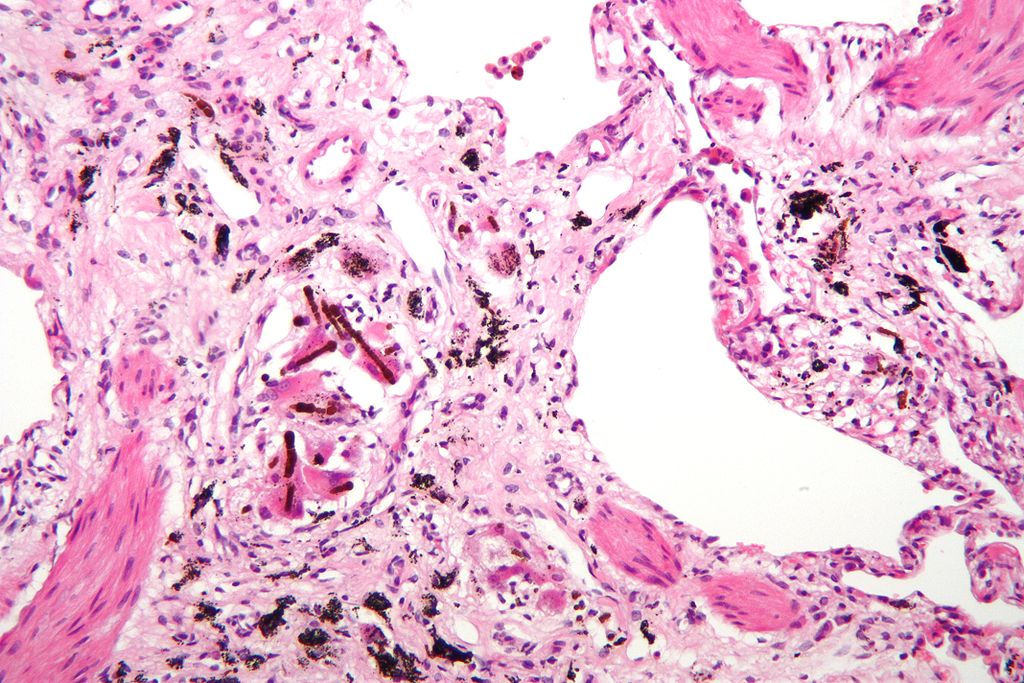
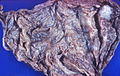
 Pneumoconiosis resulting from the inhalation of asbestos fibers.jpg Joanna Ekabua
18:32, 21 July 2020
734 × 878; 40 KB
-

-
-

-

-

-

-

-

-

-

-

-
-

-

-
-

-
-
-

-

-

-

-

-
-

-
-

-

-

-

-
-

-

-

-

-

-

-

-

-

-

-
-

-

-

-

-

-

-

-

-

-

-

-

-

-

-
-
-

-

-

-

-

-
-

-
-
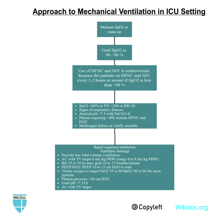
 Corel-draw-pneumonia-mechanical vent-temp.jpg Usmanaliakbar
07:22, 20 July 2020
2,273 × 1,489; 1.48 MB
-

-

-

-

-

-

-

-

-
-
-

-

-
-
-

-

-

-

-

-

-

-

-

-

-
-
-

-

-

-

-

-
-

-
-

-

-

-

-

-

-

-
-

-
-
-

-

-

-

-

-
-

-

-

-

-
-

-

-

-

-

-
-

-
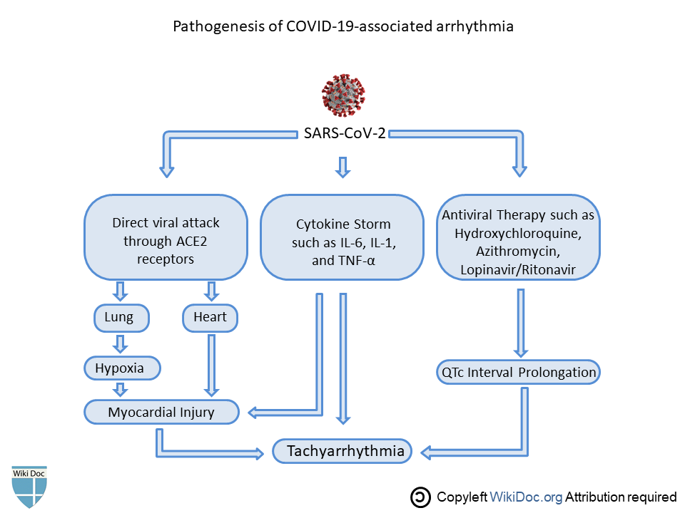
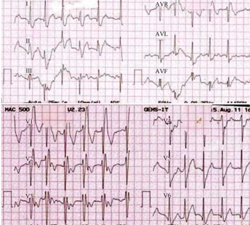
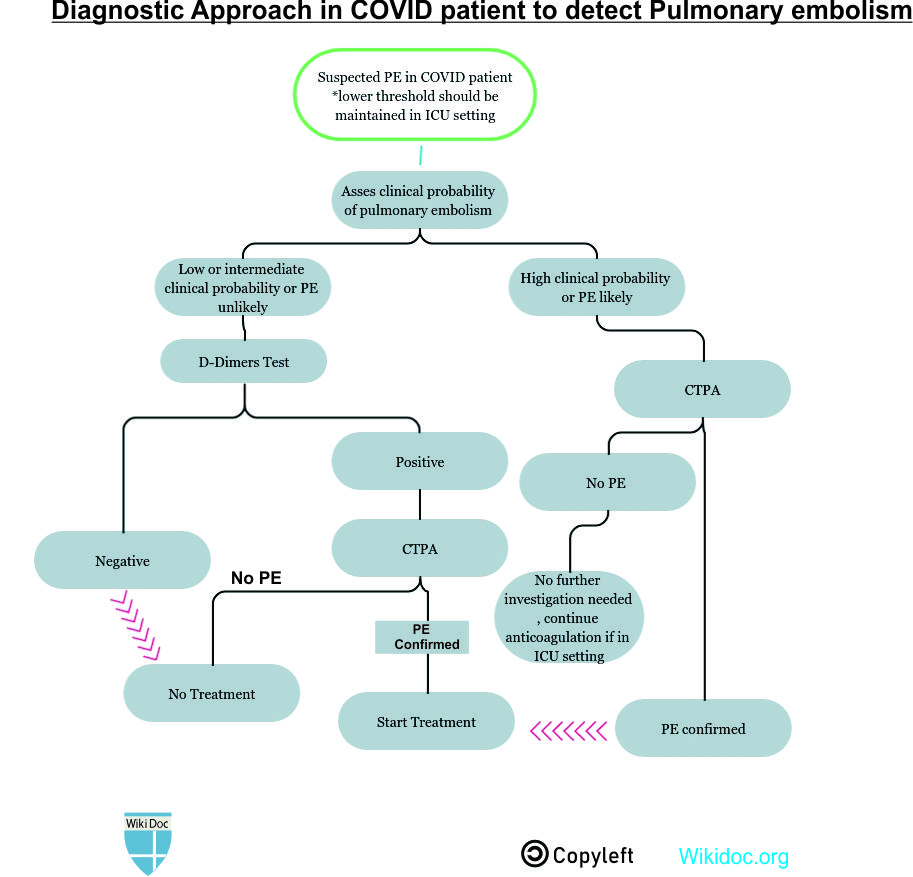
 Diagnostic approach to chest pain in COVID-19.jpg Syed rizvi
07:28, 16 July 2020
1,280 × 720; 119 KB
-

-

-

-

-
-

-
-

-

-

-

-

-

-

-

-

-

-

-

-

-

-

-

-

-
-
-

-

-

-

-

-

-

-

-

-

-

-

-

-

-

-
-

-

-

-

-

-

-

-

-

-

-

-
-

-

-

-

-

-

-
-

-
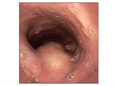
 Gastroendoscopic finiding of gastric bronchogenic cyst.jpg Joanna Ekabua
18:36, 14 July 2020
800 × 691; 110 KB
-
 Bronchoscopic findings of bronchogenicCysts Fig 2.jpg Joanna Ekabua
18:28, 14 July 2020
400 × 316; 112 KB
-

-

-

-
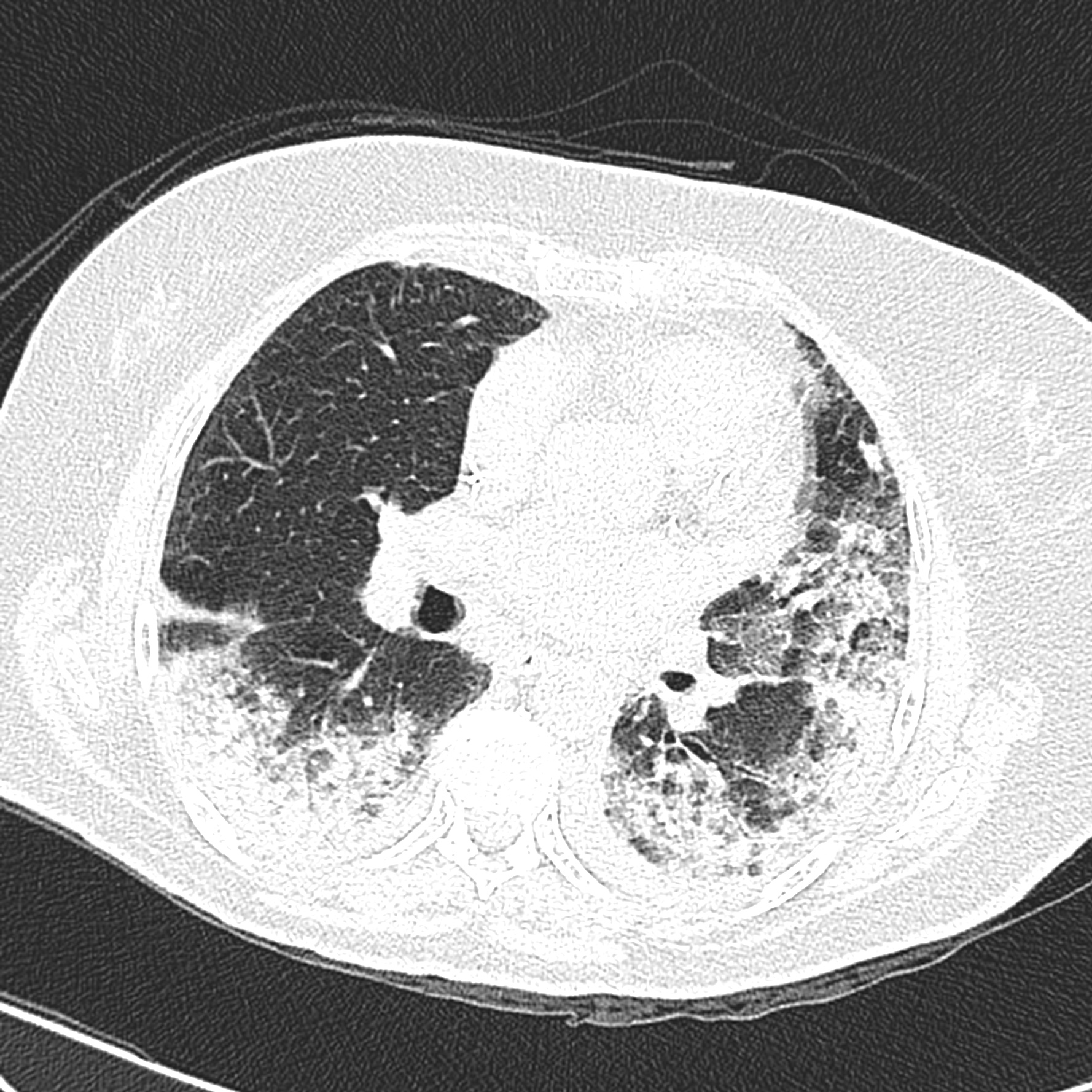
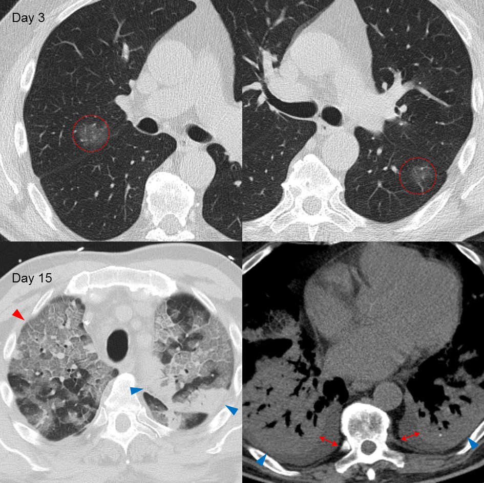
 Covid-19-pneumonia-progression-and-regression.jpg Sahar Memar Montazerin
13:27, 13 July 2020
2,836 × 2,336; 908 KB
-

-

-

-

-

-

-
-

-

-
-
-
-
-
-

-
-
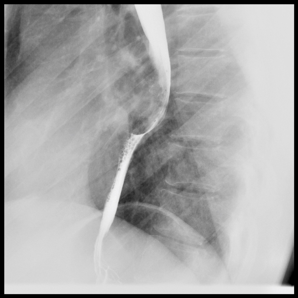
 Endoscopic view of an esophageal duplication cyst.jpg Joanna Ekabua
00:13, 12 July 2020
375 × 277; 17 KB
-
-
-
-

-

-

-

-
-

-

-

-

-

-

-
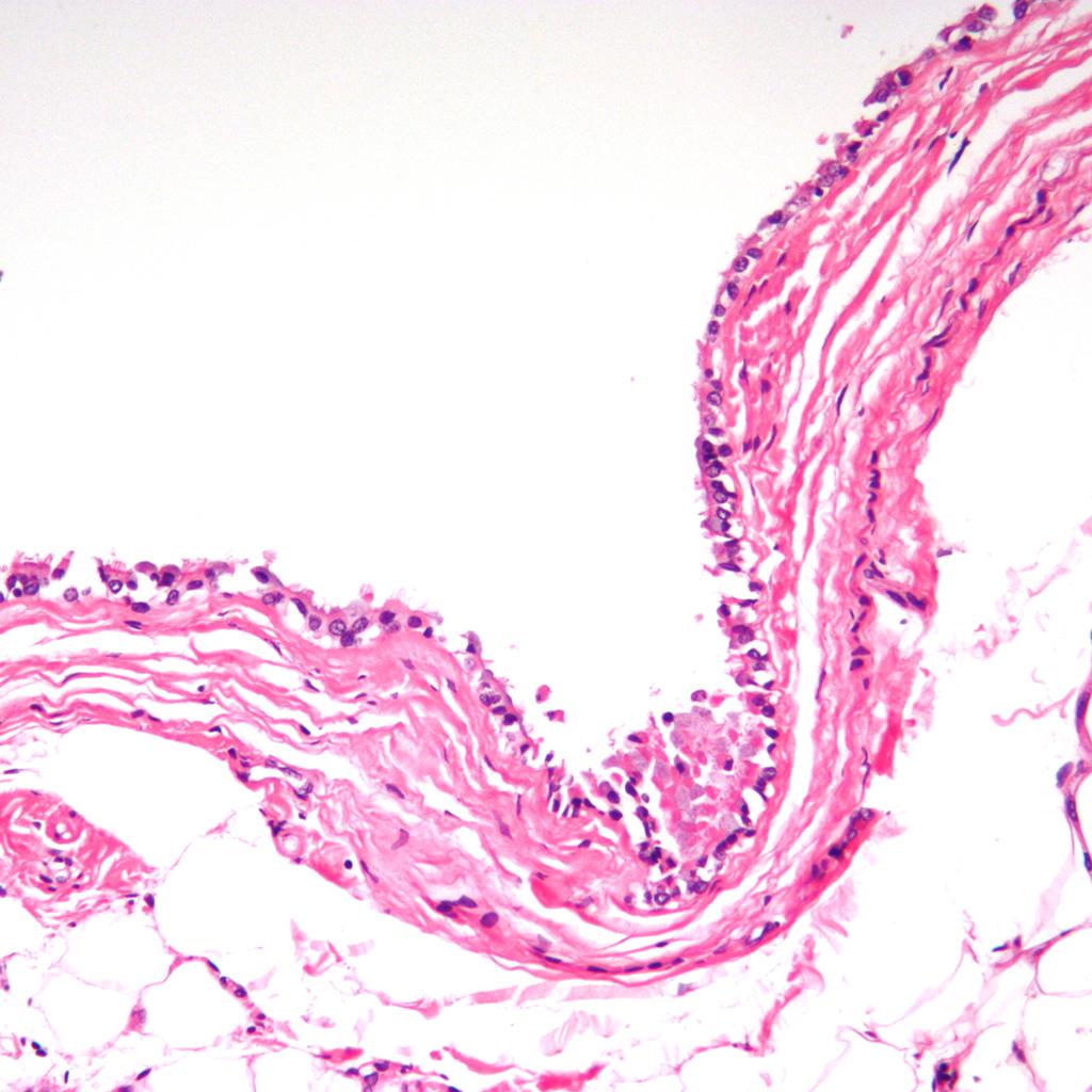
 Histologic image of bronchogenic cyst showing cilia.jpg Joanna Ekabua
22:39, 10 July 2020
1,024 × 1,024; 163 KB
-
-

-
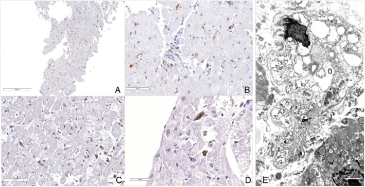
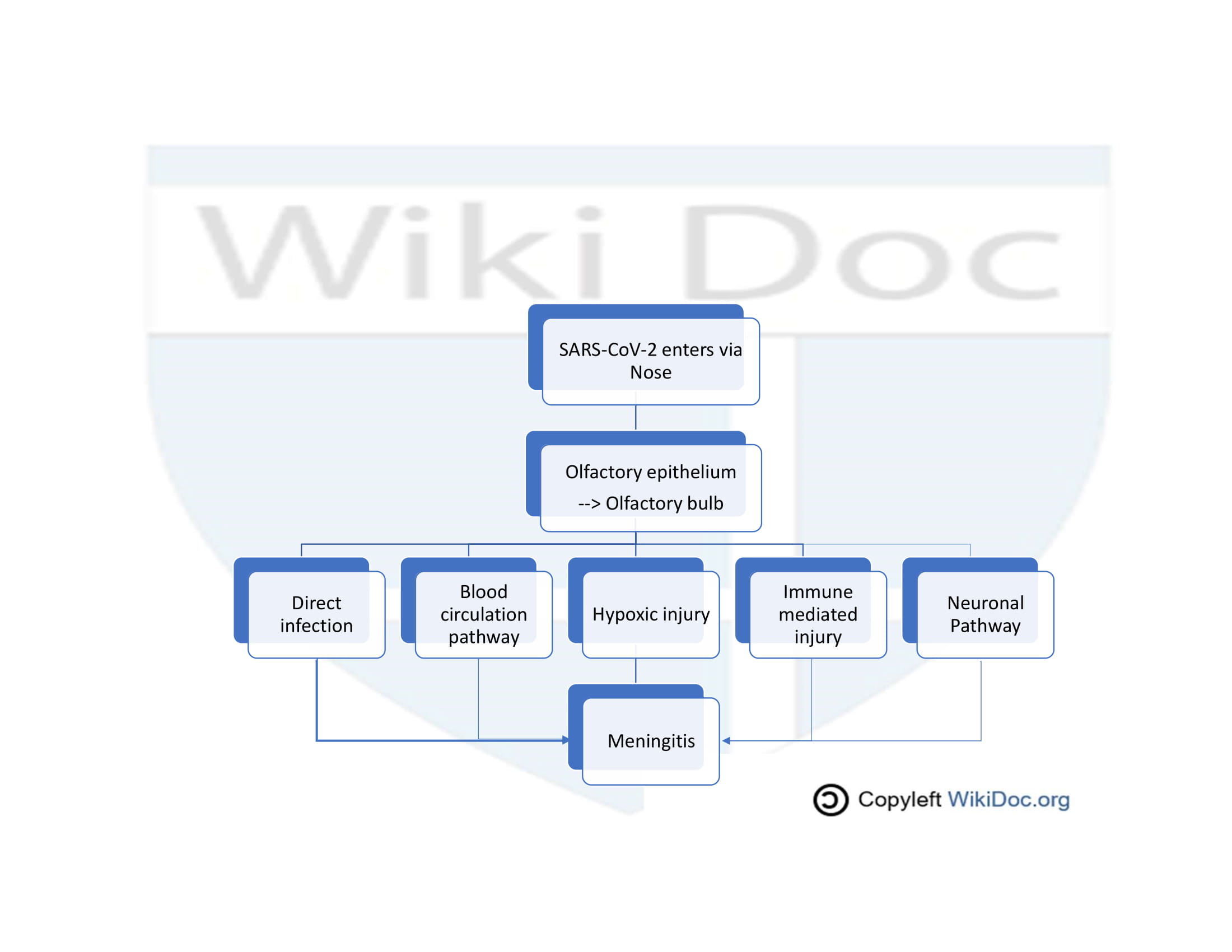
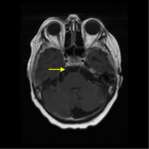 COVID-19 associated contrast enhancement CN VI.png Javaria Anwer
20:31, 10 July 2020
630 × 630; 174 KB
-

-

-
-

-

-

-
-
-

-
-
-
-
-
-
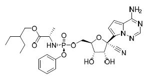
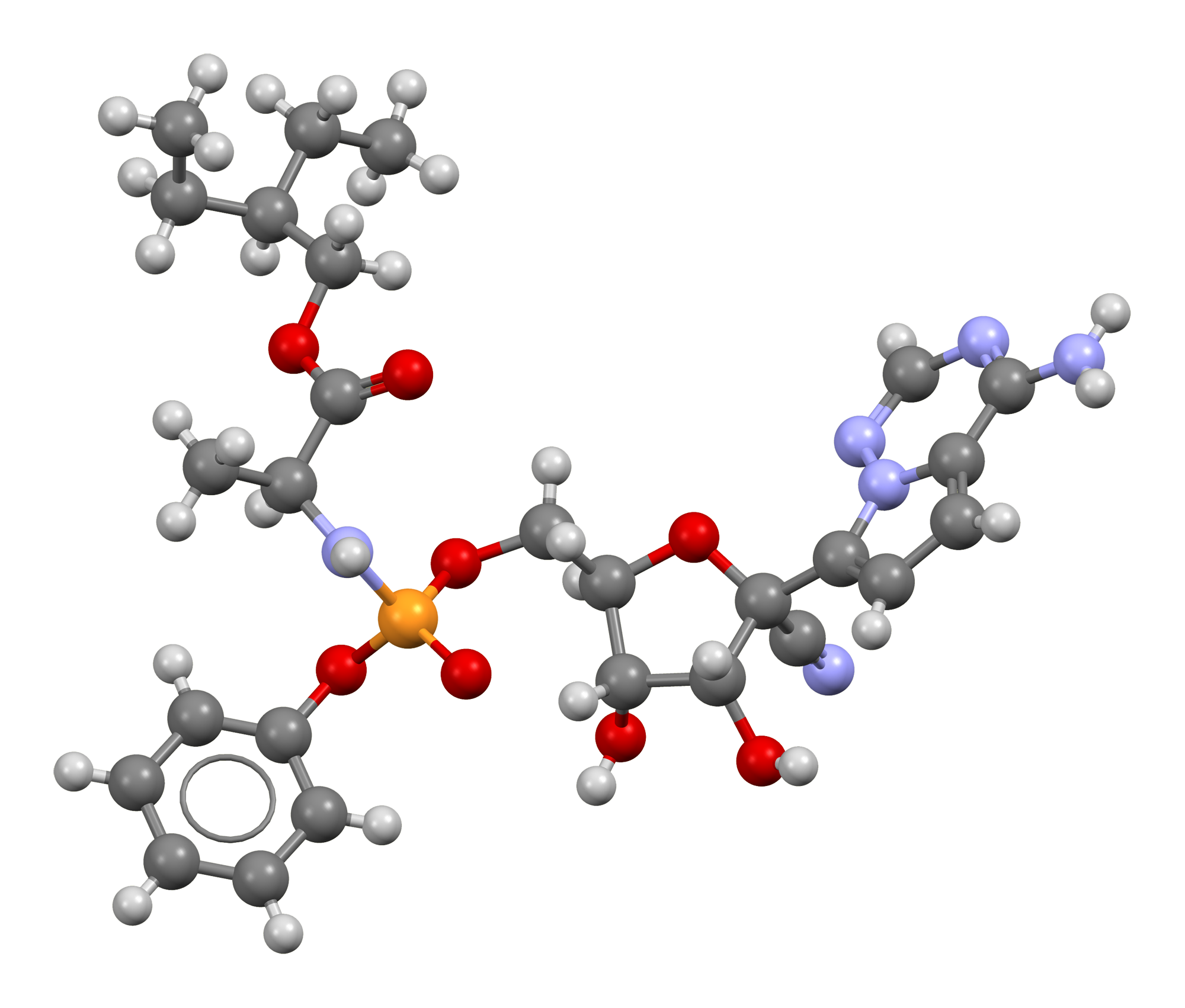 Remdesivir-from-xtal-Mercury-3D-ball-and-stick-with-bond-orders.png Gerald Chi
00:16, 3 July 2020
2,000 × 1,702; 771 KB
-

-

-
-

-
-

-

-
-

-
-
-
-

-
-

-

-

-

-
-

-

-

-
-

-

-
-

-
-

-
-

-

-
-
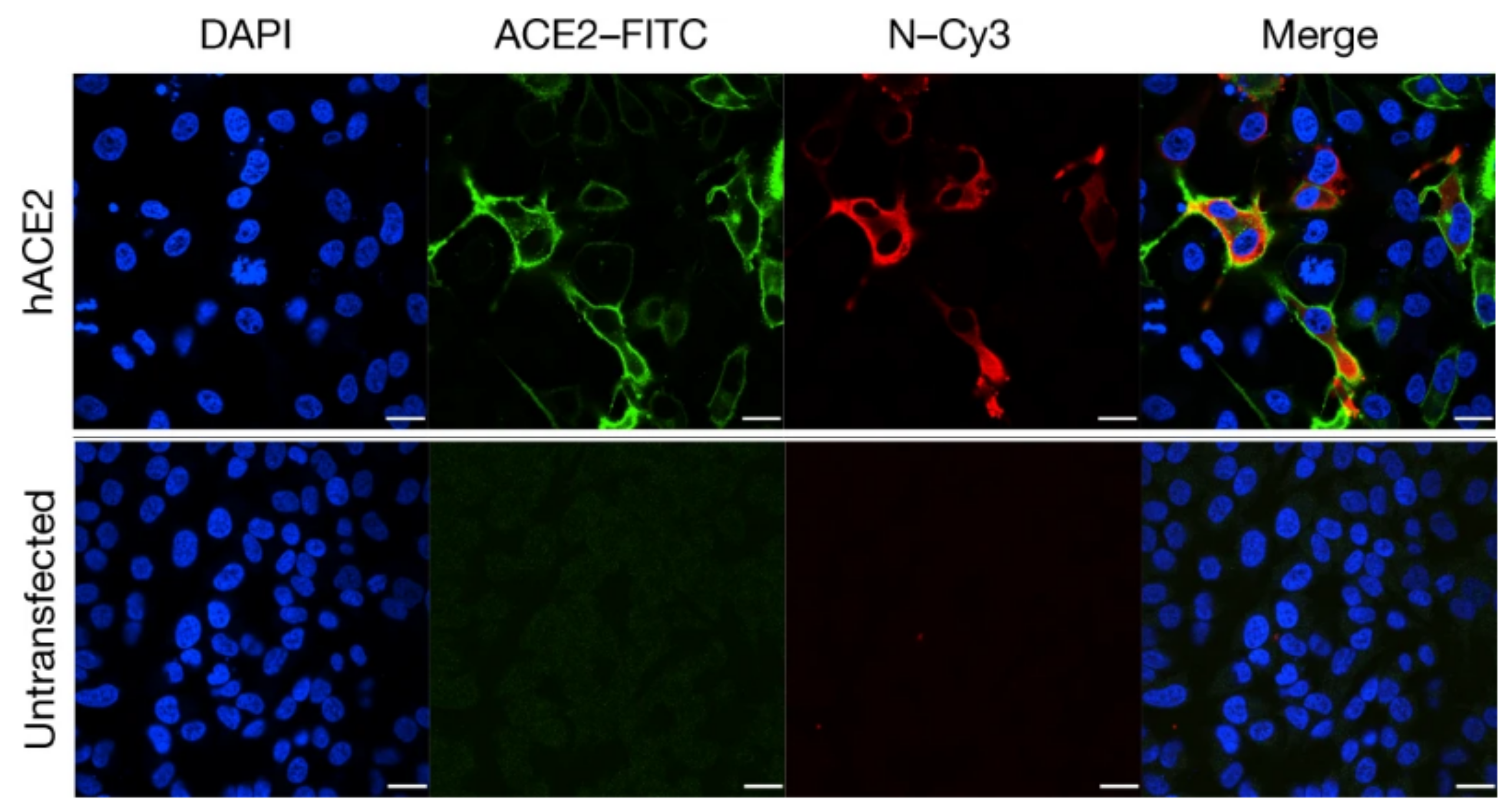 HeLa cell with ACE2 receptor SARS-COV-2.png Javaria Anwer
22:40, 27 June 2020
2,566 × 1,363; 1.77 MB
-

-
-

-

-
-

-
-
 Acute Medical Management of AV block or SND.JPG Akash Daswaney
07:42, 26 June 2020
1,730 × 1,611; 365 KB
-

-

-

-

-
-

-


.jpg)


..png)


.jpg)


.jpg)


.jpg)


.png)


.gif)


.jpg)