Gallery of new files
Jump to navigation
Jump to search
This special page shows the last uploaded files.
-
-
-

-
-
-
-
-
-
-

-
-
-
-

-
-

-
-

-

-

-

-

-

-

-

-
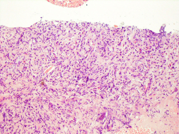
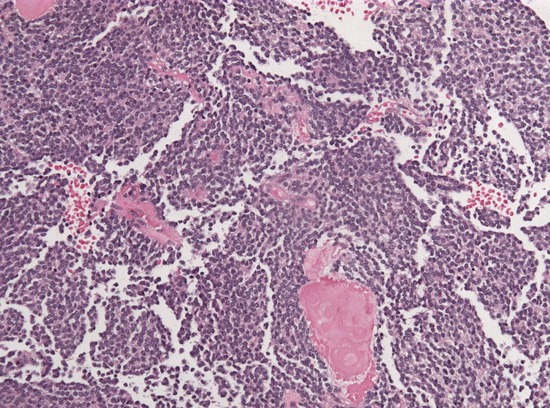
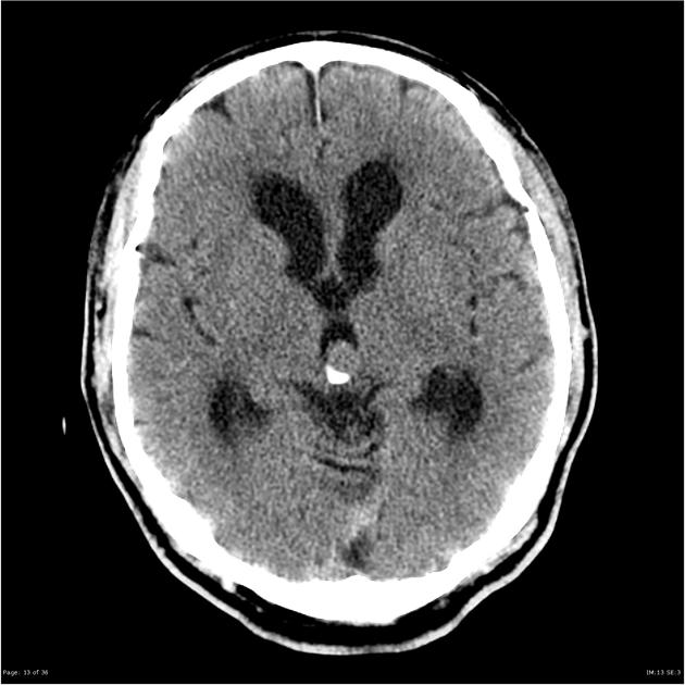
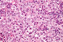
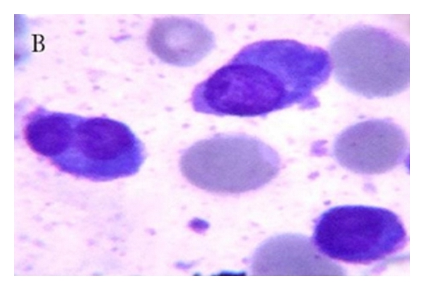
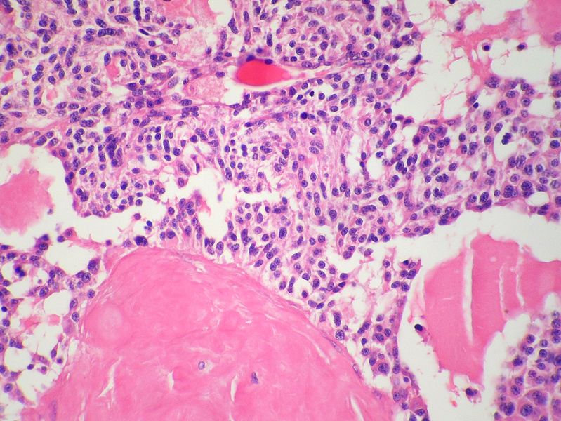
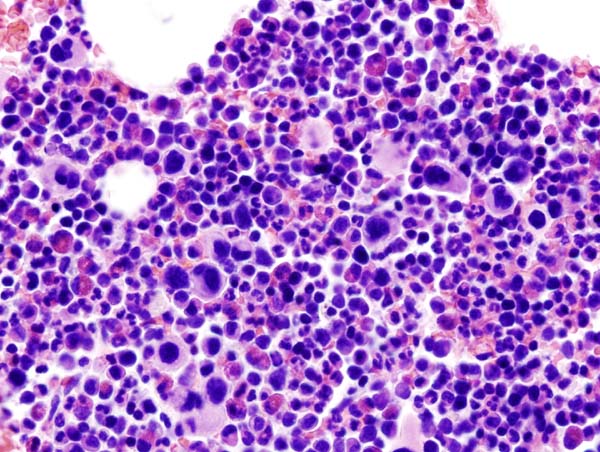
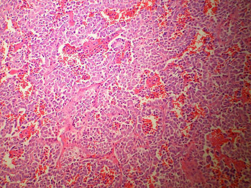
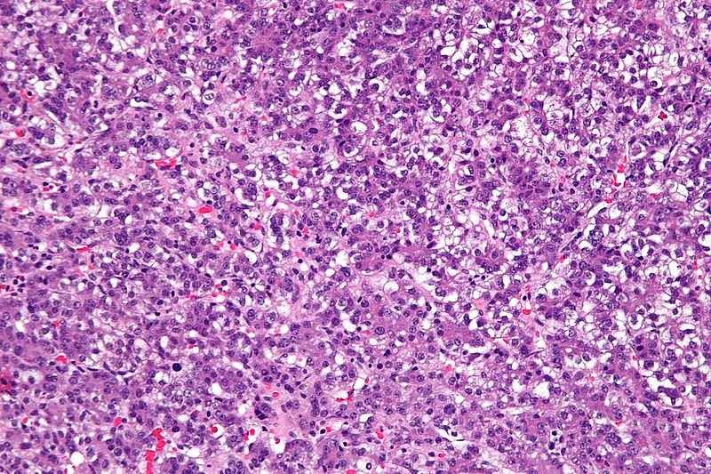
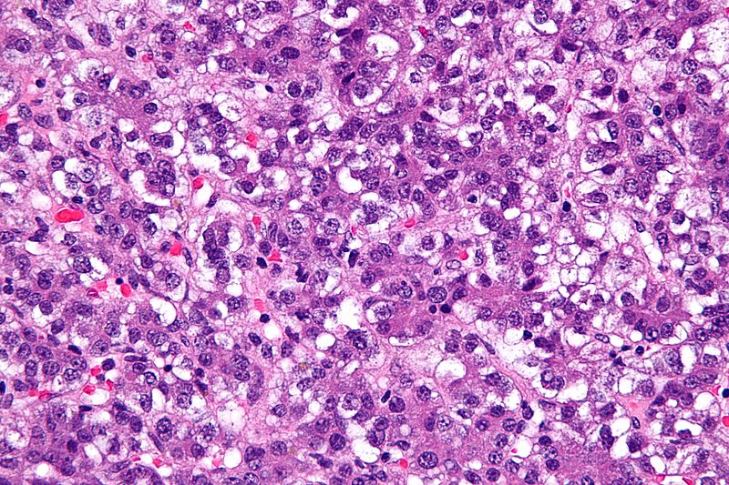
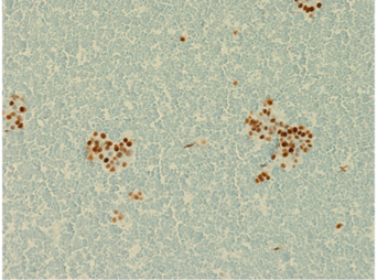
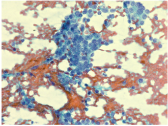
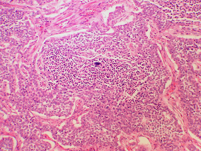
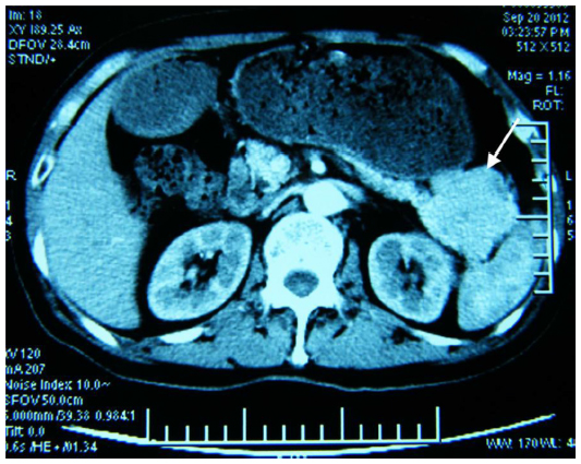
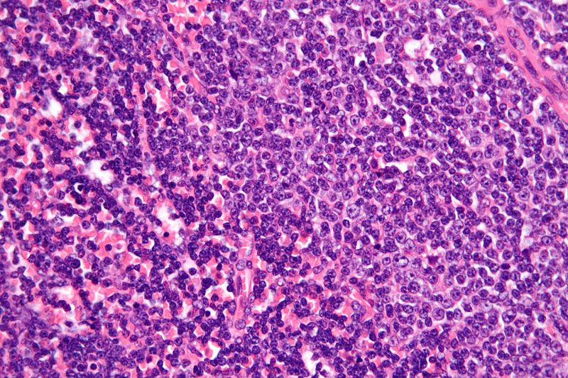
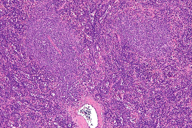
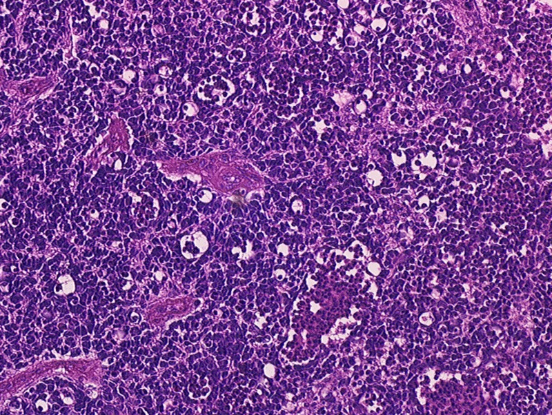
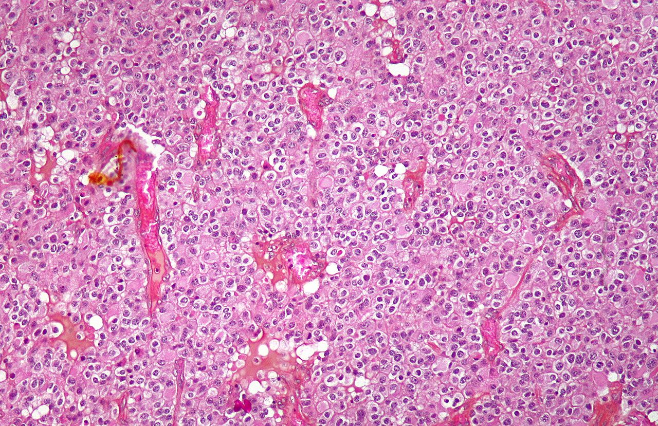
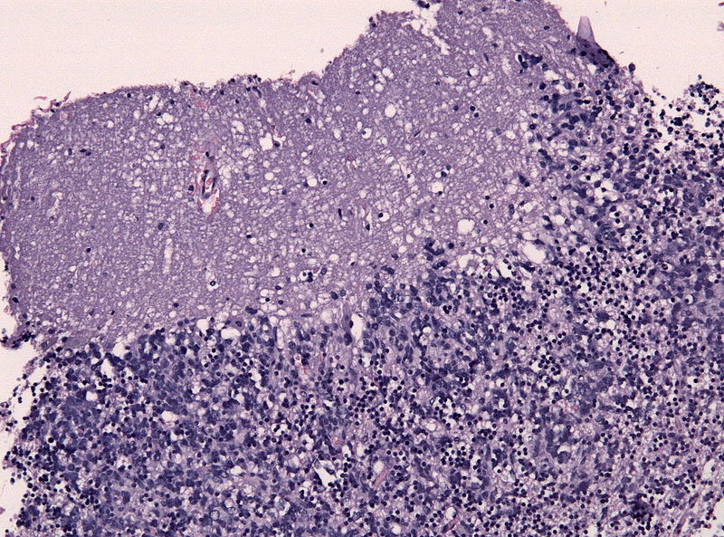
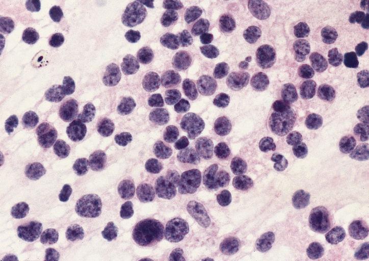
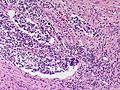
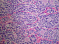
 Angioimmunoblastic T-cell lymphoma Biopsy 3.jpg Sowminya Arikapudi
16:39, 25 November 2015
600 × 450; 429 KB
-
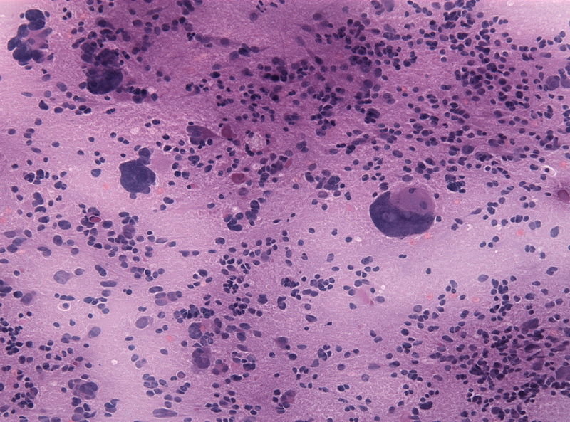
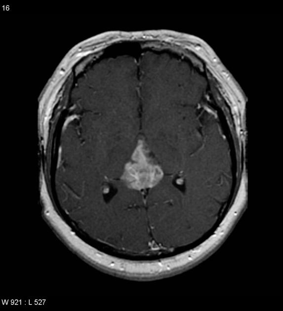
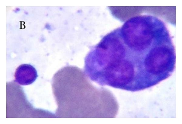
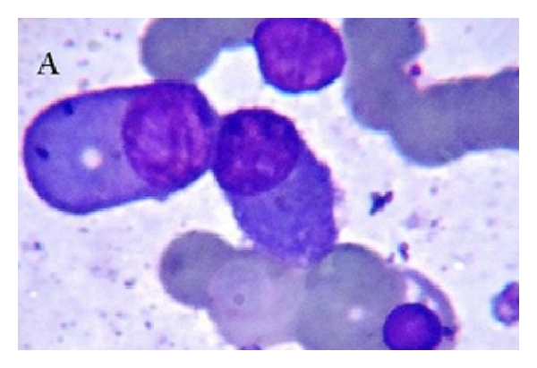
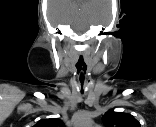
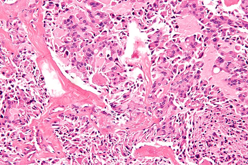
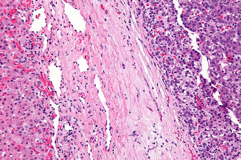
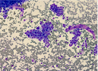
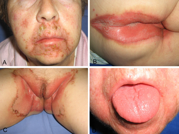
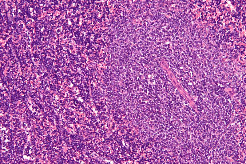
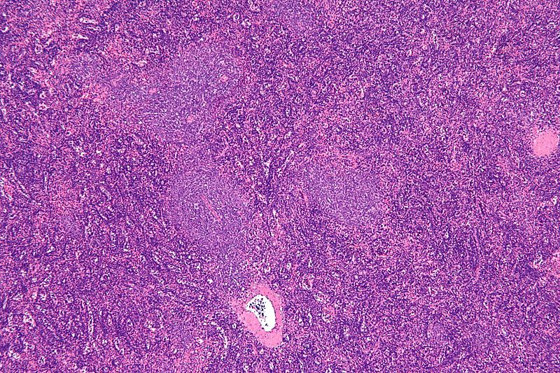
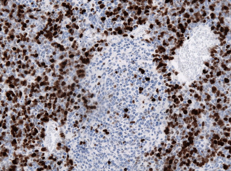
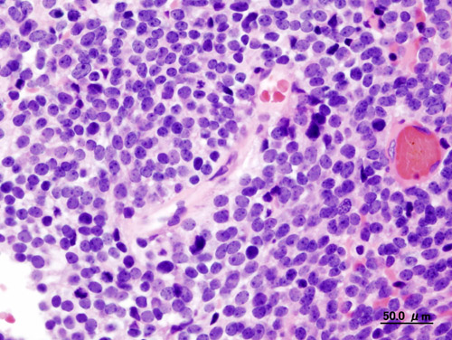
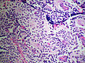
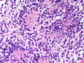
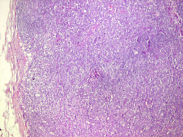 Angioimmunoblastic T-cell lymphoma Biopsy 2.jpg Sowminya Arikapudi
16:38, 25 November 2015
600 × 450; 460 KB
-
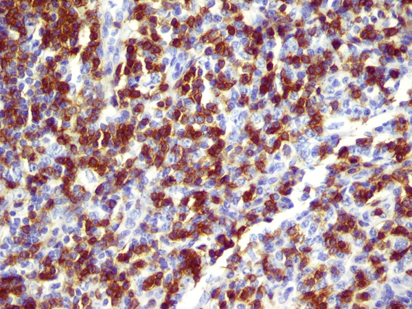
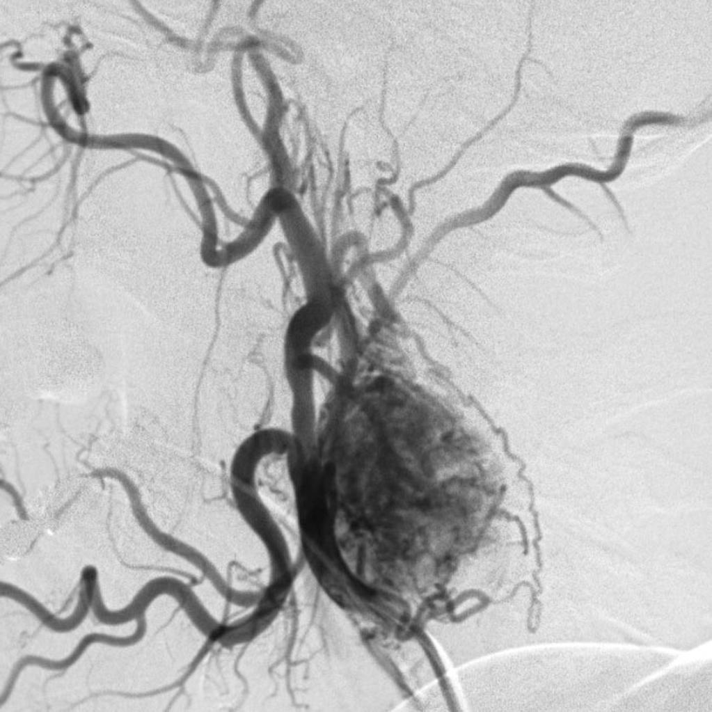
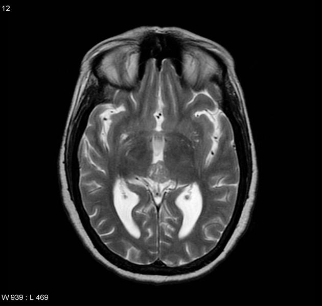
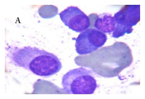
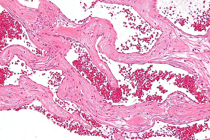
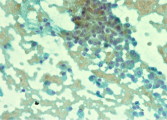
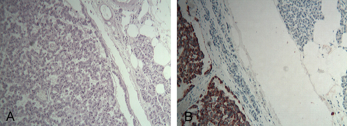
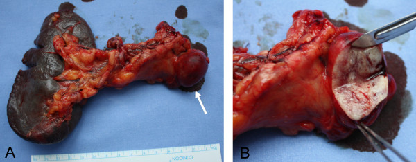
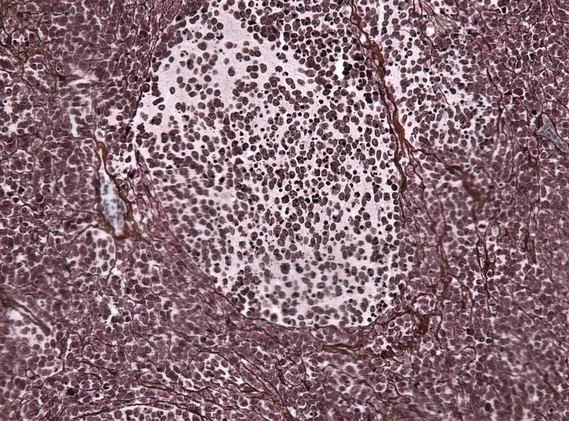
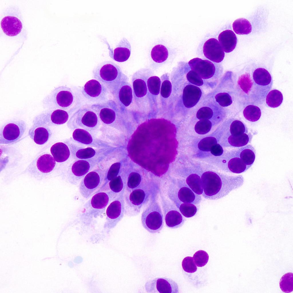
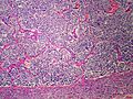
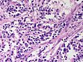
 Angioimmunoblastic T-cell lymphoma Biopsy 1.jpg Sowminya Arikapudi
16:37, 25 November 2015
600 × 450; 377 KB
-
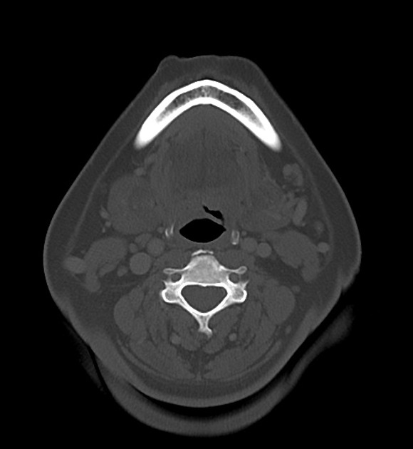
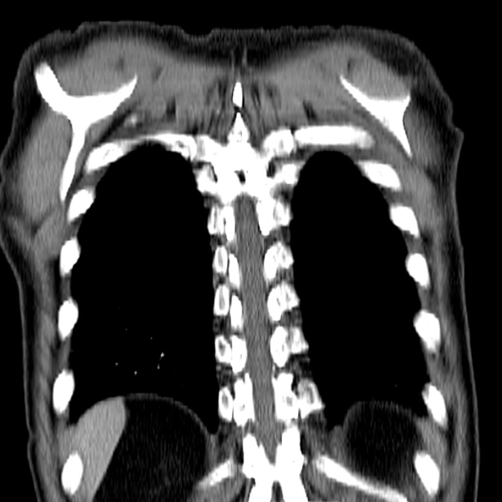
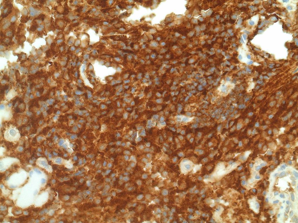
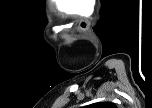
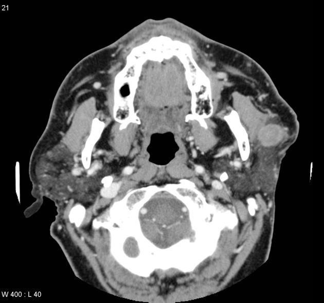
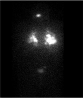
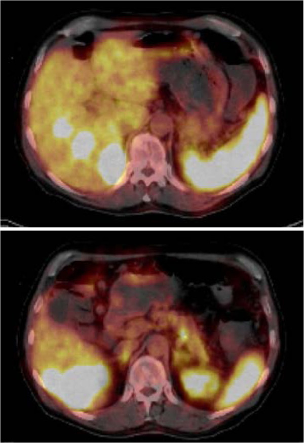
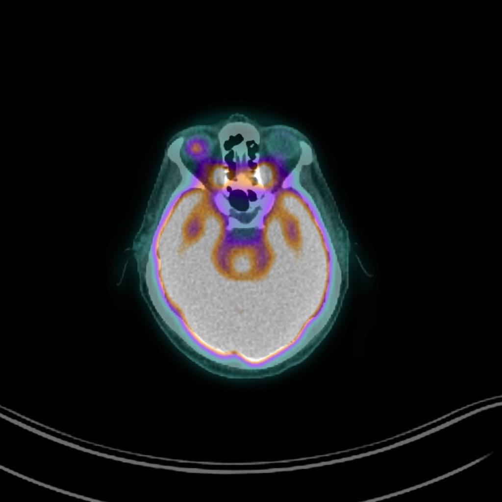
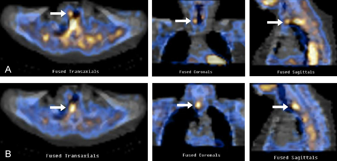
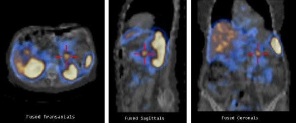
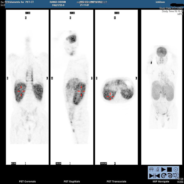 Angioimmunoblastic T-cell lymphoma PET Scan.jpg Sowminya Arikapudi
16:18, 25 November 2015
600 × 600; 126 KB
-
-
-
-

-
-
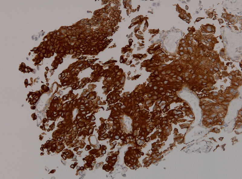
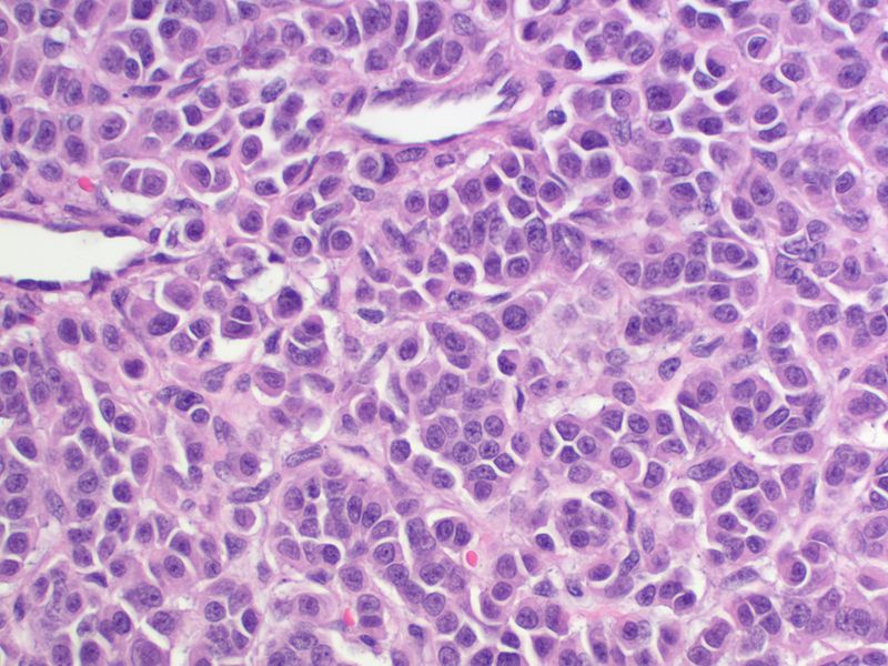
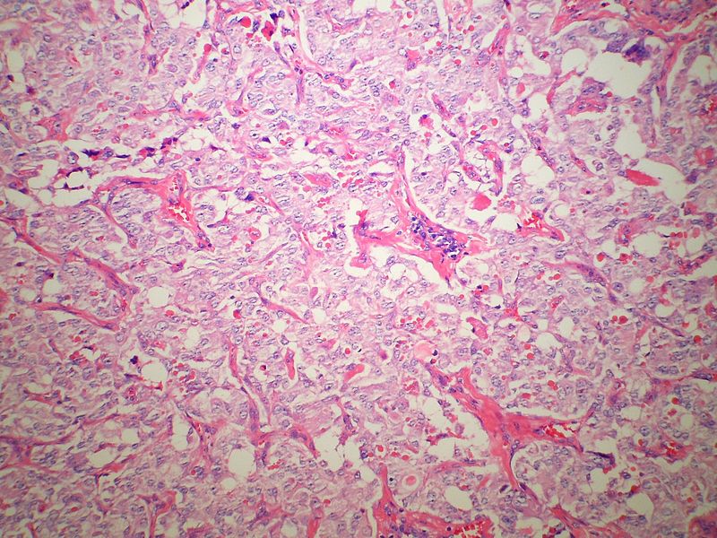
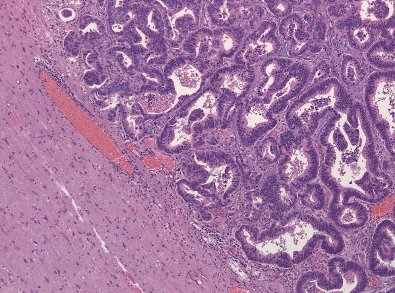
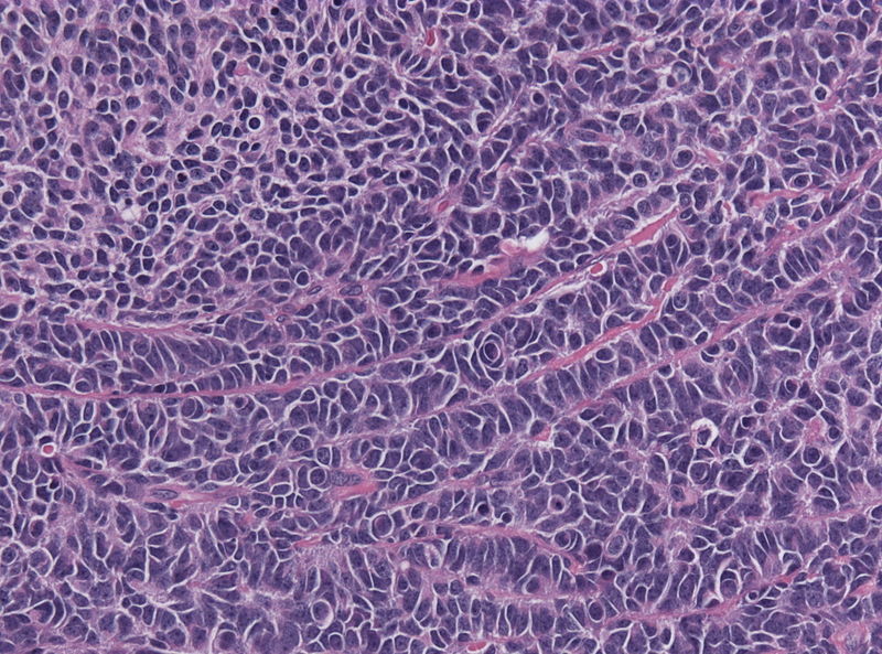
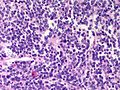
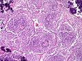
 800px-PTPR papillary tumor pinealis HE microscopic image 2.jpg Sujit Routray
18:37, 24 November 2015
800 × 593; 130 KB
-
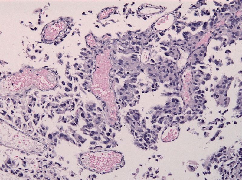
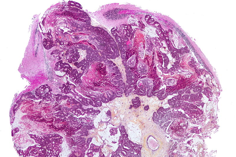
 Papillary Tumor of the Pineal Region microscopic image 1.PNG Sujit Routray
18:24, 24 November 2015
883 × 736; 390 KB
-
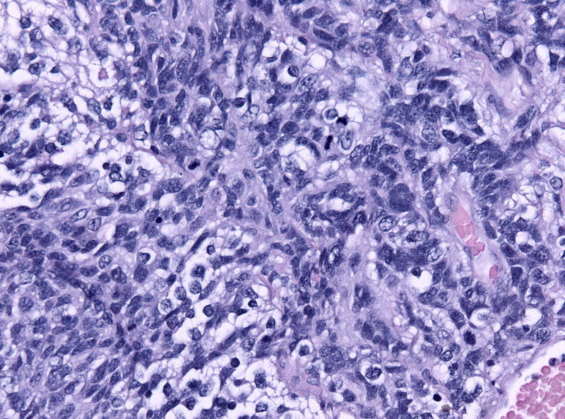
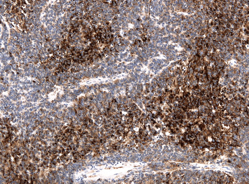
 Pineal parenchymal tumor with intermediate differentiation microscopic 4.jpg Sujit Routray
18:32, 23 November 2015
800 × 593; 90 KB
-
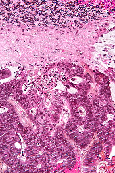
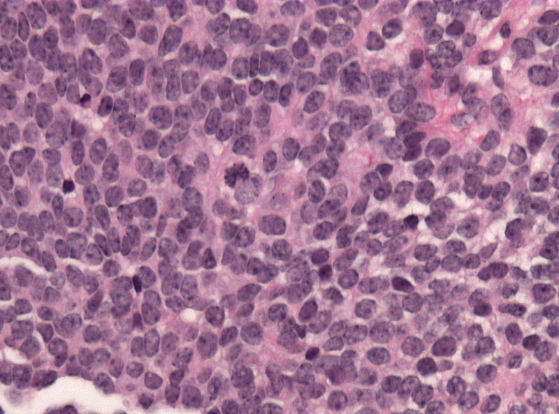 Pineal parenchymal tumor with intermediate differentiation microscopic 3.jpg Sujit Routray
18:27, 23 November 2015
800 × 593; 103 KB
-
 Pineal parenchymal tumor with intermediate differentiation microscopic 2.jpg Sujit Routray
18:23, 23 November 2015
800 × 593; 176 KB
-
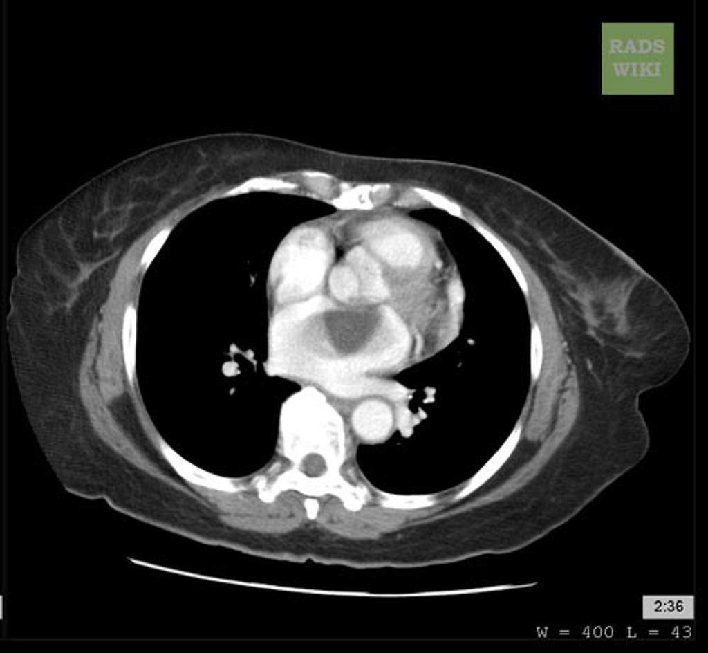
 Left-hilar-mass-small-cell-lung-cancer-2.png.jpeg Mirdula Sharma
18:17, 23 November 2015
1,024 × 793; 170 KB
-
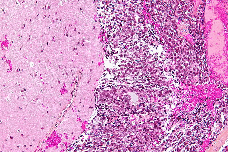
 Pineal parenchymal tumor with intermediate differentiation microscopic 1.jpg Sujit Routray
18:15, 23 November 2015
800 × 593; 131 KB
-

-
-
-
-
-

-

-

-
-
-
-

-
-
-
-
-
-

-
-
-
-
-
-
-

-
-
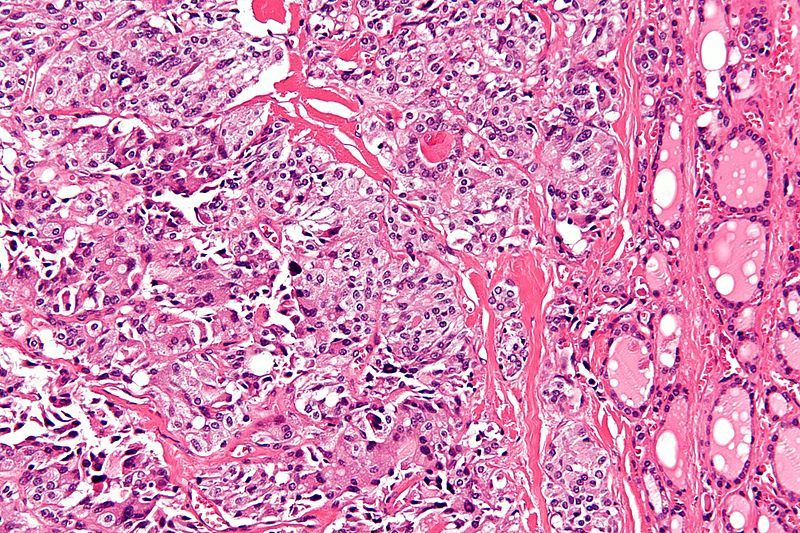
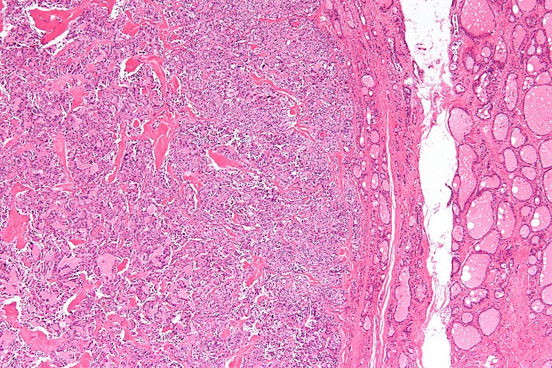
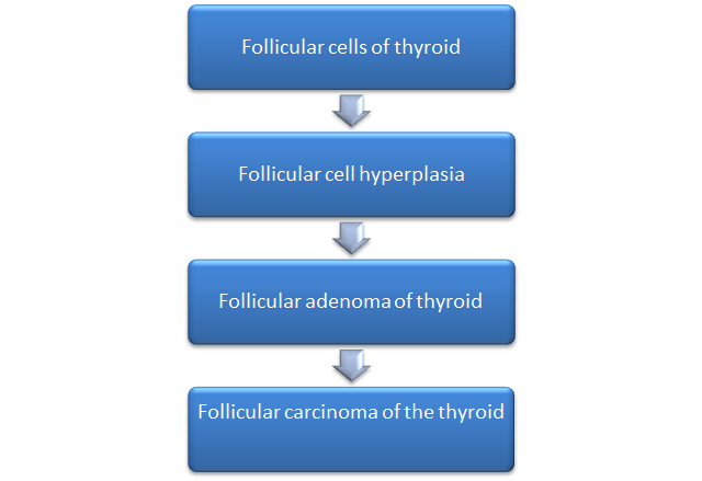
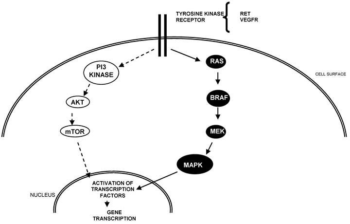
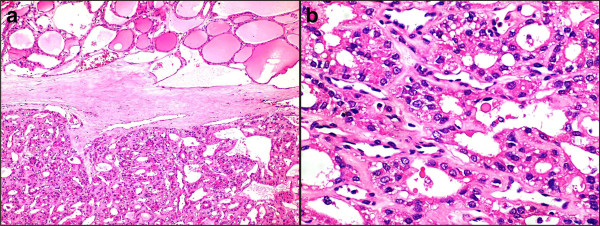
 Follicular thyroid carcinoma pathogenesis 2.0.jpg Ammu Susheela
21:14, 17 November 2015
960 × 720; 40 KB
-
-
-
-
-

-
-
-
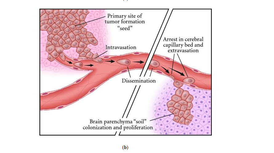
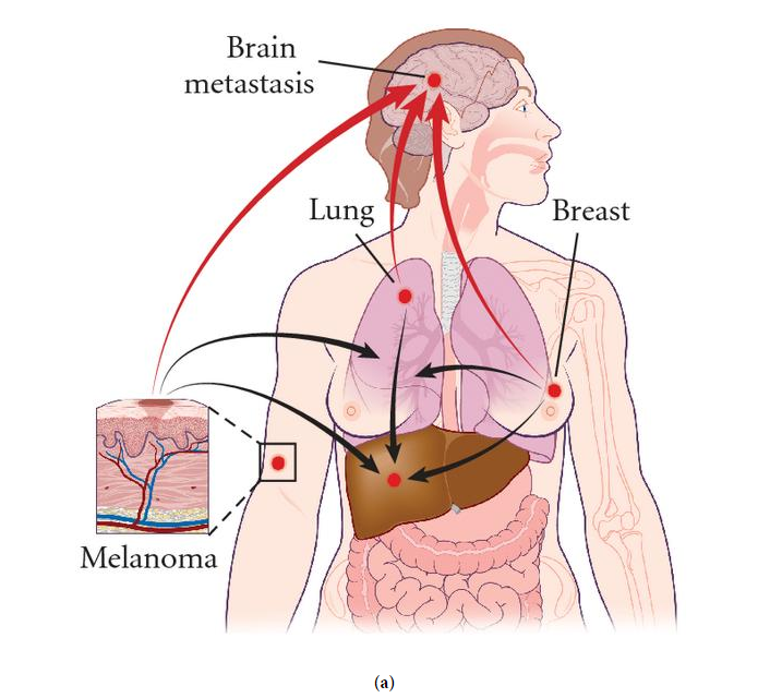
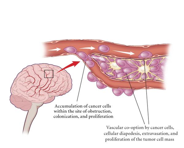 Colonization of metastatic tumor cells in the brain.jpg Sujit Routray
22:40, 13 November 2015
600 × 485; 33 KB
-
-
-
-
-
-
-
-
-
-
-
-
-
-
-
-
-
-
-
-
-
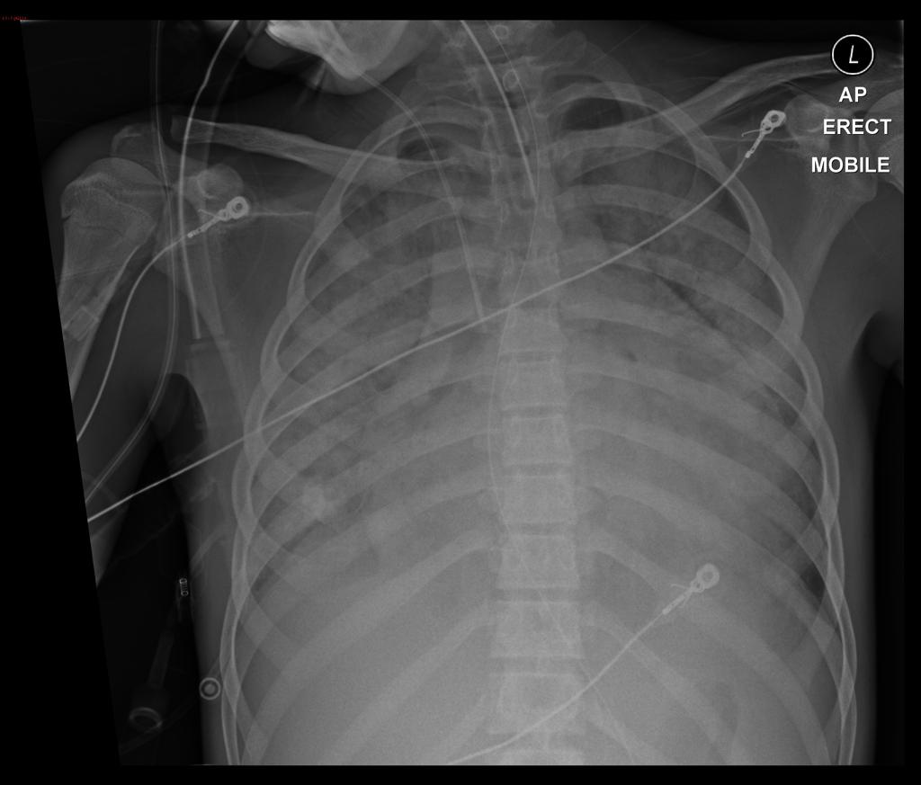
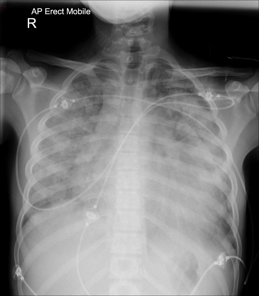
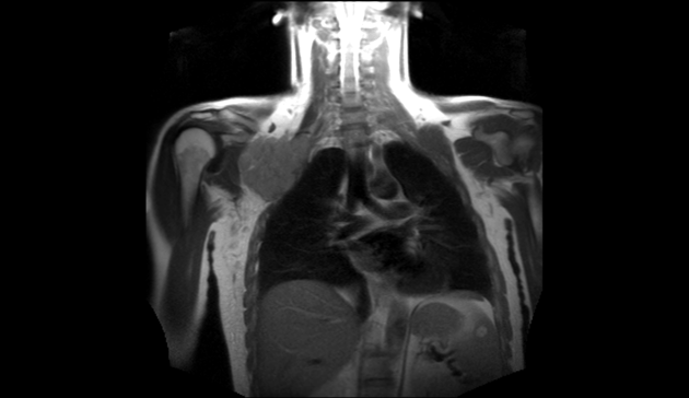
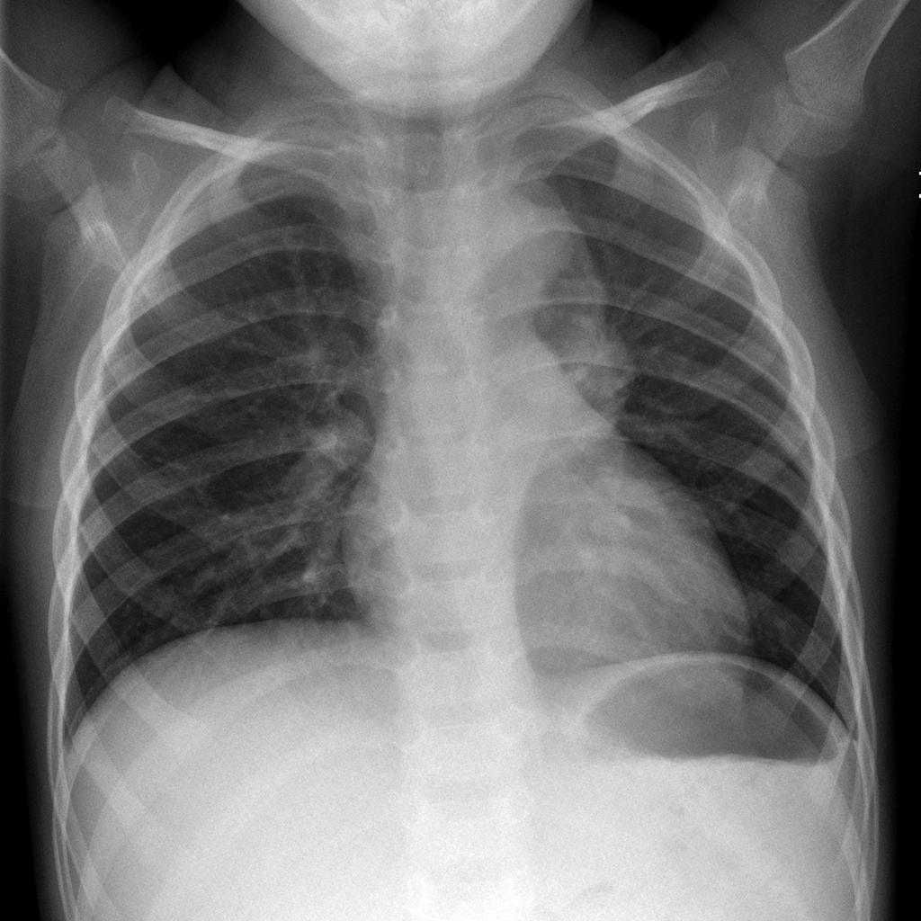
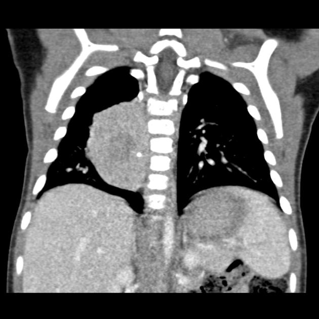
 Chest X ray of metastatic bronchogenic carcinoma.jpg Sujit Routray
16:24, 13 November 2015
1,024 × 1,011; 82 KB
-
-
-
-
-
-
-
-
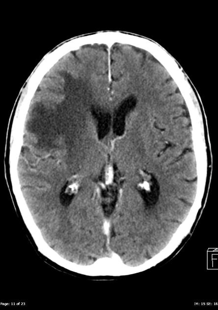
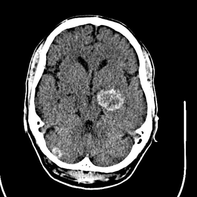
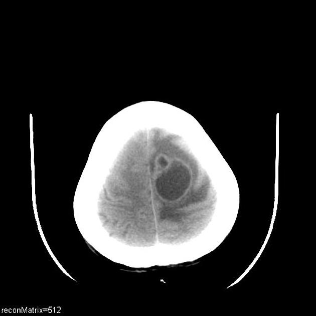
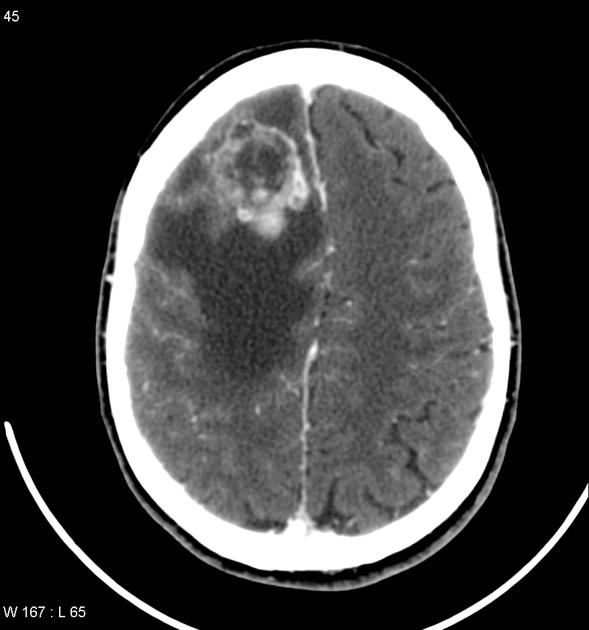
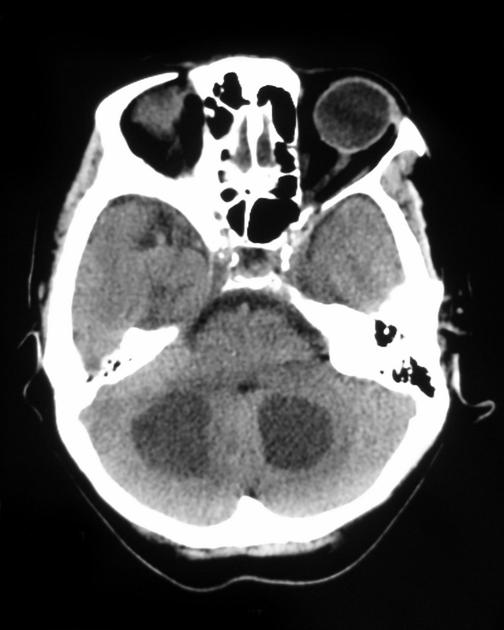
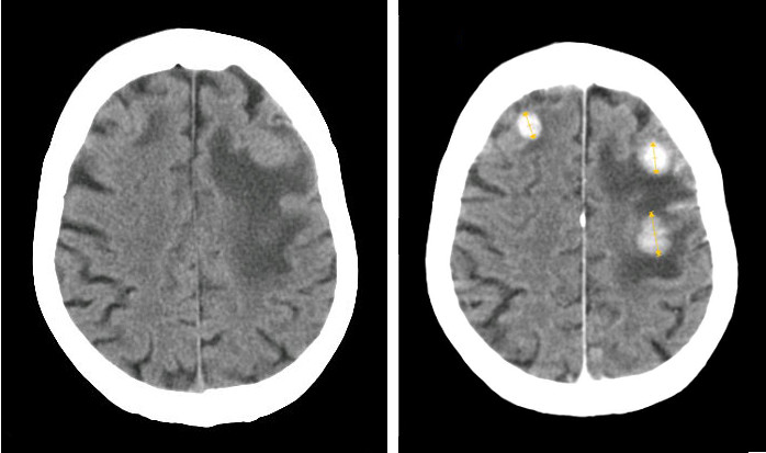
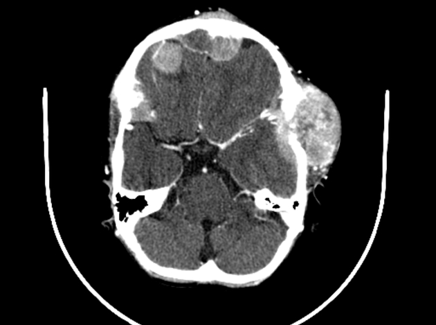
 CT scan of brain metastasis 7 postcontrast.jpg Sujit Routray
15:09, 13 November 2015
630 × 630; 17 KB
-
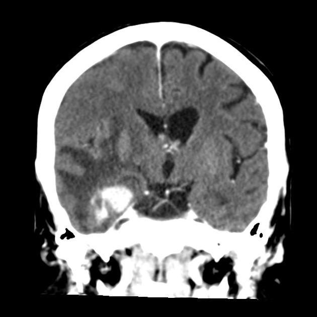
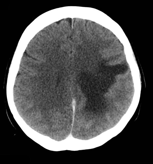
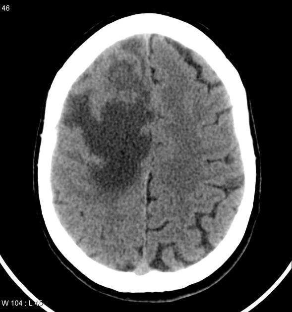
 CT scan of brain metastasis 6 noncontrast.jpg Sujit Routray
15:07, 13 November 2015
630 × 630; 18 KB
-
-
-
-
-
-
 Thyroid MedullaryCarcinoma Amyloid HP PA.JPG Ammu Susheela
21:17, 12 November 2015
799 × 600; 114 KB
-

-
 Thyroid MedullaryCarcinoma SmallCellVariant HP CTR (3).jpg Ammu Susheela
21:12, 12 November 2015
800 × 600; 111 KB
-
 Thyroid MedullaryCarcinoma SmallCellVariant MP CTR.jpg Ammu Susheela
21:09, 12 November 2015
800 × 600; 184 KB
-
 Thyroid MedullaryCarcinoma SmallCell HP PA.JPG Ammu Susheela
21:07, 12 November 2015
799 × 600; 171 KB
-
 Thyroid MedullaryCarcinoma SpindleCell LP2 PA.JPG Ammu Susheela
21:05, 12 November 2015
766 × 600; 110 KB
-
 Thyroid MedullaryCarcinoma SpindleCell MP PA.JPG Ammu Susheela
21:03, 12 November 2015
799 × 600; 143 KB
-
 Thyroid MedullaryCarcinoma SpindleCell LP PA.JPG Ammu Susheela
20:56, 12 November 2015
799 × 600; 163 KB
-
 Thyroid MedullaryCarcinoma Comedonecrosis HP CTR.jpg Ammu Susheela
20:51, 12 November 2015
800 × 600; 88 KB
-
 Thyroid MedullaryCarcinoma Comedonecrosis LP2 CTR.jpg Ammu Susheela
20:47, 12 November 2015
800 × 600; 127 KB
-
 Thyroid MedullaryCarcinoma Amyloid MP PA.JPG Ammu Susheela
20:42, 12 November 2015
799 × 600; 139 KB
-
 Thyroid MedullaryCarcinoma Amyloid MP4 PA.JPG Ammu Susheela
20:38, 12 November 2015
799 × 600; 126 KB
-

-
-
-
-
-
-
 Thyroid MedullaryCarcinoma Amyloid MP2 PA.JPG Ammu Susheela
20:15, 12 November 2015
799 × 600; 136 KB
-

-
 Thyroid MedullaryCarcinoma Amyloid MP3 PA.JPG Ammu Susheela
20:13, 12 November 2015
799 × 600; 136 KB
-
 800px-Thyroid MedullaryCarcinoma Amyloid RBWH.JPG Ammu Susheela
20:11, 12 November 2015
800 × 594; 154 KB
-

-

-
-
-
-
-
-
-
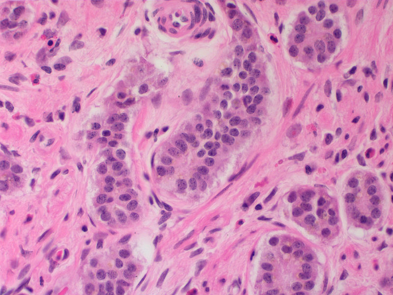
 Medullary thyroid carcinoma - 2 - high mag.jpg Ammu Susheela
19:42, 12 November 2015
800 × 533; 158 KB
-

-
-
-

-
-
-

-
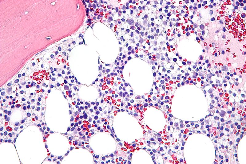
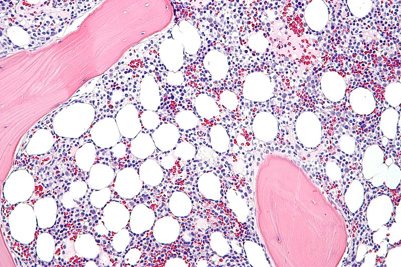
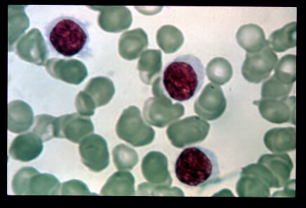
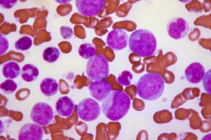
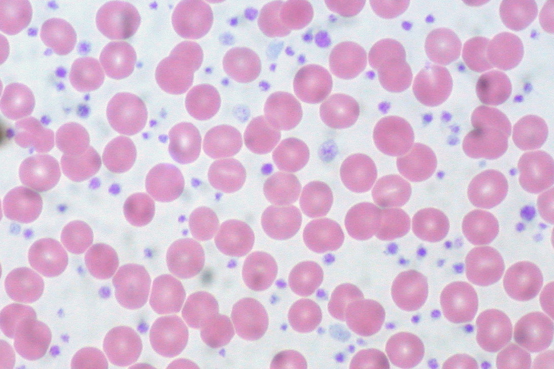 Essential Thrombocythemia, Peripheral Blood.jpg Soujanya Thummathati
19:02, 12 November 2015
1,092 × 727; 234 KB
-
-
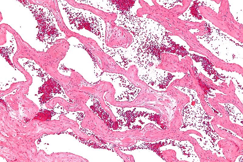
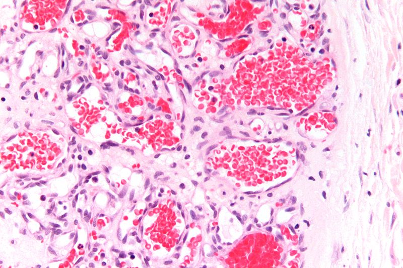 Capillary hemangioma very high magnification.jpg Nawal Muazam
18:48, 12 November 2015
800 × 533; 106 KB
-
-
 Capillary hemangioma intermediate magnification.jpg Nawal Muazam
18:37, 12 November 2015
800 × 533; 176 KB
-
-
-

-
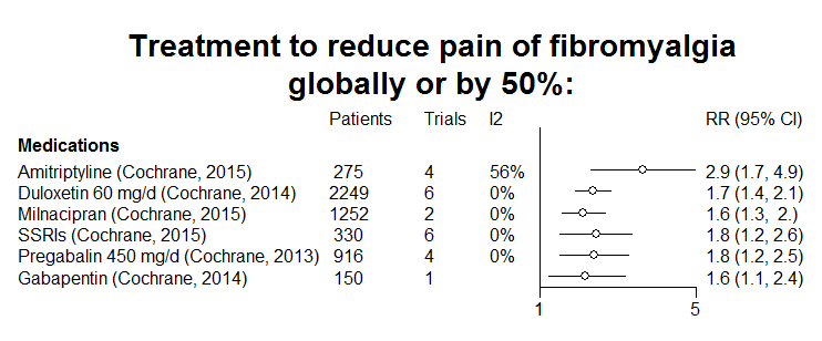
 Blobbogram of treatment of chronic low back pain.png Badgettrg
06:55, 12 November 2015
967 × 858; 63 KB
-
-

-
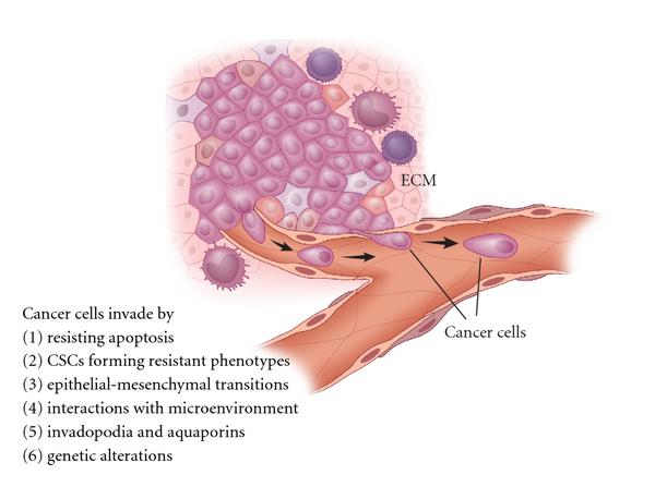
 Schematics of the process of metastasis 2.PNG Sujit Routray
14:27, 11 November 2015
890 × 500; 408 KB
-
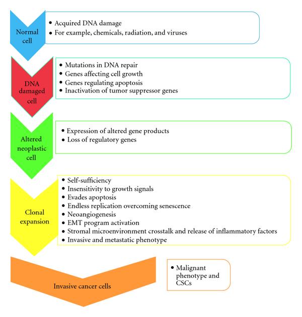
 Schematics of the process of metastasis 1.PNG Sujit Routray
14:14, 11 November 2015
717 × 640; 446 KB
-
 1600px-Medulloblastoma with rosettes.jpg Maria Villarreal
19:28, 10 November 2015
1,600 × 1,186; 403 KB
-
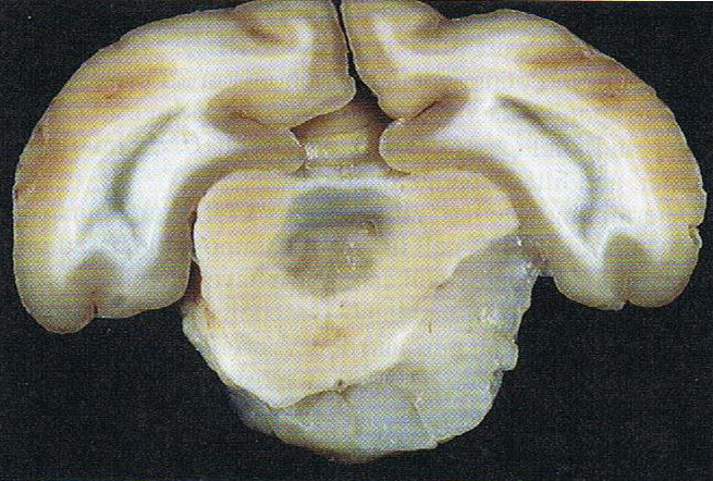
 Cerebellar medulloblastoma (1) in adult.jpg Maria Villarreal
19:17, 10 November 2015
500 × 376; 109 KB
-
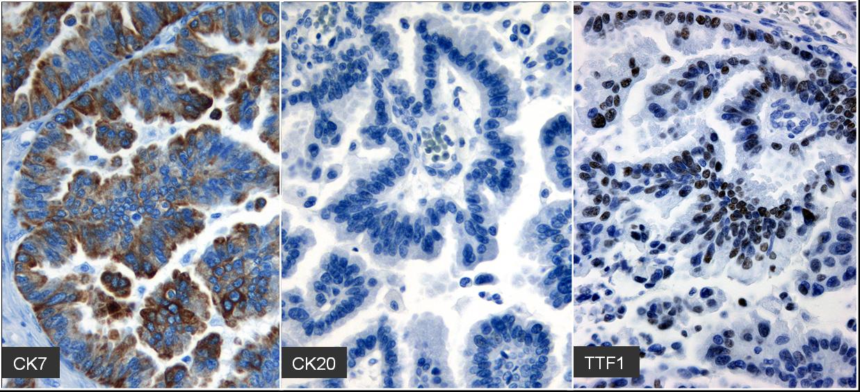 Immunohistochemistry of brain metastasis.jpg Sujit Routray
18:58, 10 November 2015
1,237 × 565; 162 KB
-
-
-
-
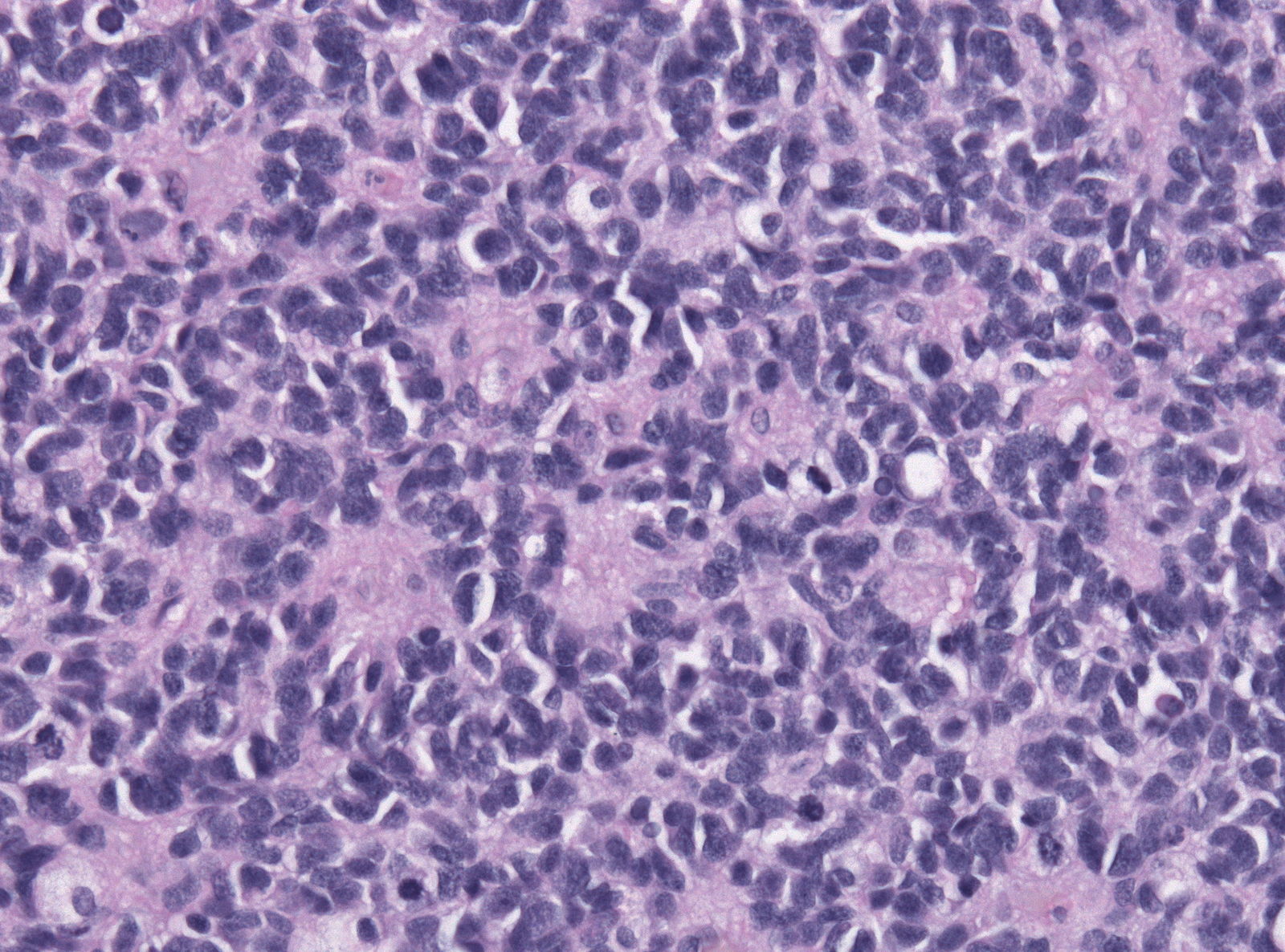
 Metastatic adenocarcinoma - cerebellum - high mag.jpg Sujit Routray
15:46, 10 November 2015
400 × 600; 117 KB
-
 Metastatic adenocarcinoma - cerebellum - very low mag.jpg Sujit Routray
15:37, 10 November 2015
800 × 533; 155 KB
-
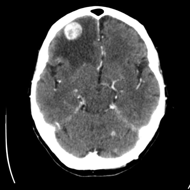
 THYROID PAPILLARY CARCINOMA METASTATIC TO BRAIN.jpg Sujit Routray
15:15, 10 November 2015
789 × 512; 267 KB
-
-
-
 Mucoepidermoid carcinoma (2) HE stain (1).jpg Simrat Sarai
06:06, 10 November 2015
600 × 452; 384 KB
-
 800px-Salivary duct carcinoma -a- low mag.jpg Simrat Sarai
05:58, 10 November 2015
800 × 533; 201 KB
-
 800px-Epithelial-myoepithelial carcinoma - high mag (1).jpg Simrat Sarai
05:57, 10 November 2015
800 × 533; 158 KB
-
 800px-Basal cell adenocarcinoma - parotid gland - high mag.jpg Simrat Sarai
05:55, 10 November 2015
800 × 533; 159 KB
-
 800px-Adenoid cystic carcinoma - high mag.jpg Simrat Sarai
05:54, 10 November 2015
800 × 533; 183 KB
-
 796px-Papillary cystadenoma lymphomatosum2.jpg Simrat Sarai
05:51, 10 November 2015
796 × 600; 165 KB
-
 412px-Polymorphous low-grade adenocarcinoma - very low mag.jpg Simrat Sarai
05:41, 10 November 2015
412 × 599; 89 KB
-
 Whole brain external beam radiotherapy 1.PNG Sujit Routray
22:05, 9 November 2015
1,274 × 684; 310 KB
-

-
-
 Diagram showing after surgery for medullary thyroid cancer with the central lymph nodes and the thyroid gland removed CRUK 092.png Ammu Susheela
20:36, 9 November 2015
278 × 258; 45 KB
-
-

-
-

-
-
-
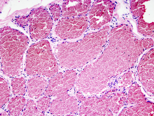
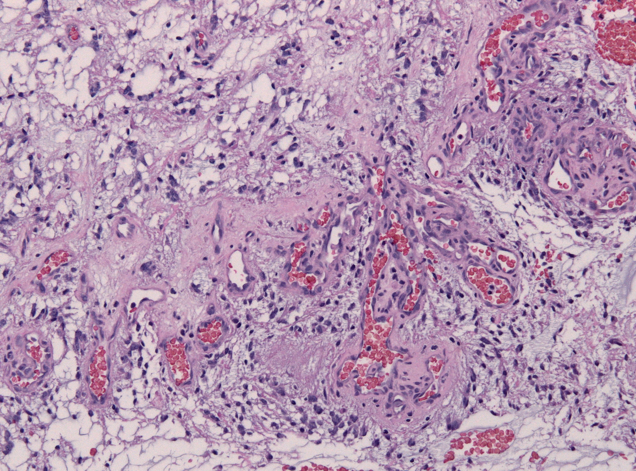
 Cavernous liver hemangioma high magnification.jpg Nawal Muazam
17:13, 7 November 2015
800 × 533; 178 KB
-
-

-
 Subependymal giant cell astrocytoma CT scan 1.jpg Sujit Routray
22:19, 4 November 2015
495 × 630; 45 KB
-
-
-
-

-
-
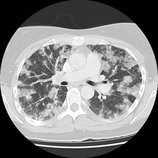
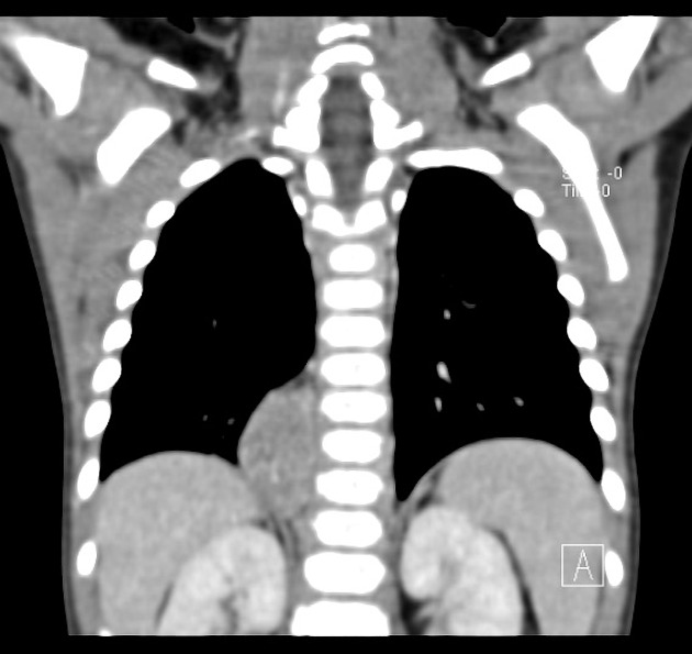
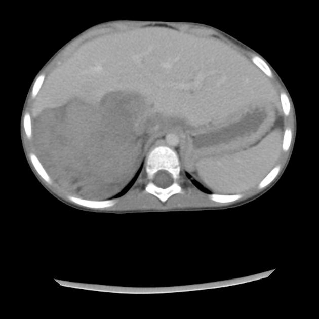
 Miliary metastasis from papillary thyroid cancer CT.jpeg Ammu Susheela
16:28, 3 November 2015
630 × 622; 68 KB
-
-

-
-

-

-
-
-

-

-
 Papillary thyroid cancer with miliary metastasis.jpg Ammu Susheela
16:12, 3 November 2015
442 × 442; 9 KB
-
-
 Papillary thyroid cancer with nodal metastasis.jpeg Ammu Susheela
16:02, 3 November 2015
504 × 630; 36 KB
-

-
-
-
 Pilocytic astrocytoma microscopic compact and loose patterns.PNG Sujit Routray
15:16, 3 November 2015
325 × 246; 194 KB
-
 Thyroid papillary carcinoma histopathology (4).jpg Ammu Susheela
15:04, 3 November 2015
500 × 376; 59 KB
-
 Thyroid papillary carcinoma histopathology (3).jpg Ammu Susheela
15:03, 3 November 2015
500 × 376; 55 KB
-
 Thyroid papillary carcinoma histopathology (2).jpg Ammu Susheela
15:01, 3 November 2015
1,024 × 771; 322 KB
-
 Thyroid papillary carcinoma histopatholgy (1).jpg Ammu Susheela
14:57, 3 November 2015
1,024 × 771; 489 KB
-
 Papillary thyroid carcinoma tall cell var intermed mag.jpg Ammu Susheela
14:56, 3 November 2015
1,024 × 683; 229 KB
-
 -Papillary thyroid carcinoma tall cell var high mag.jpg Ammu Susheela
14:55, 3 November 2015
1,024 × 683; 211 KB
-
 -Lymph node with papillary thyroid carcinoma.jpg Ammu Susheela
14:53, 3 November 2015
800 × 769; 153 KB
-
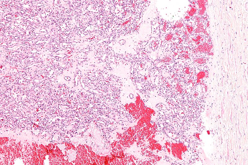
 Subependymal giant cell astrocytoma microscopic appearance 1.jpg Sujit Routray
01:08, 3 November 2015
2,048 × 1,536; 421 KB
-
 Subependymal giant cell astrocytoma microscopic appearance 4.jpg Sujit Routray
01:02, 3 November 2015
2,080 × 1,542; 858 KB
-
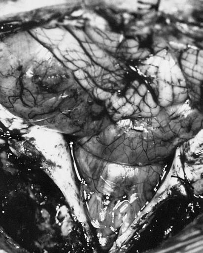
 Subependymal giant cell astrocytoma gross appearance 1.jpg Sujit Routray
00:33, 3 November 2015
717 × 512; 221 KB
-
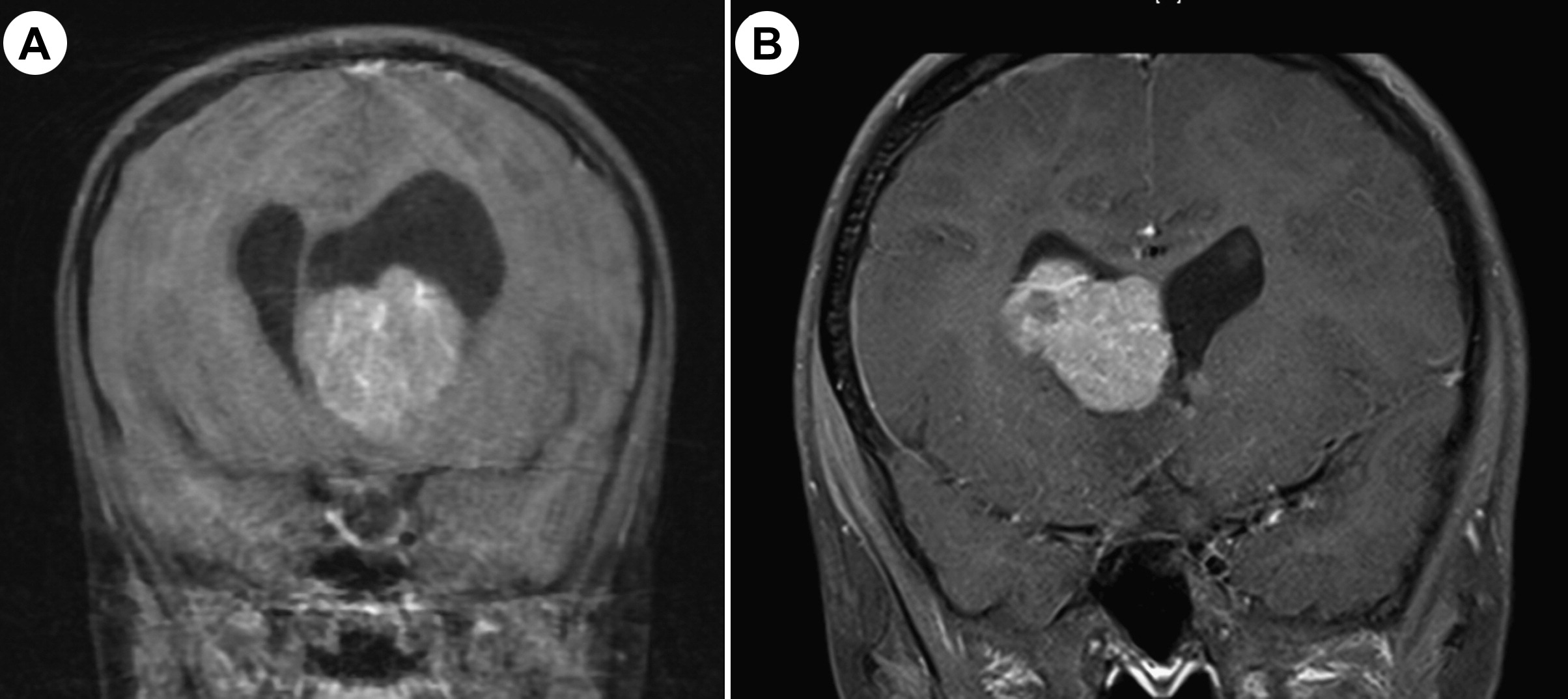
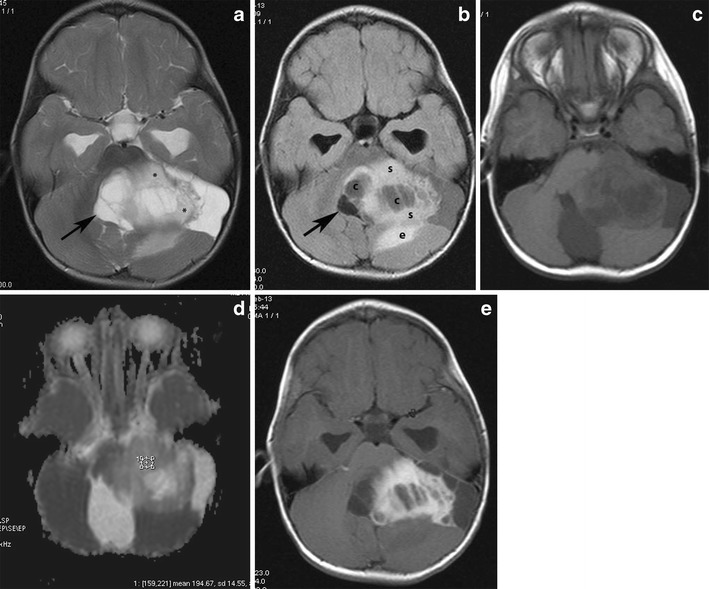
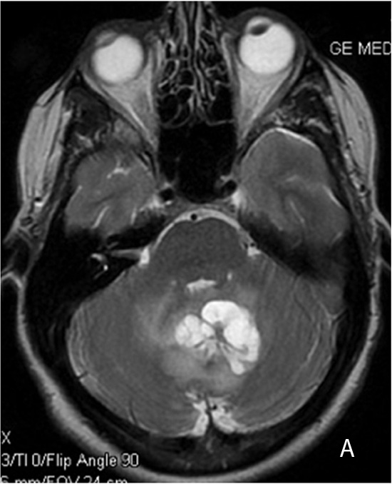
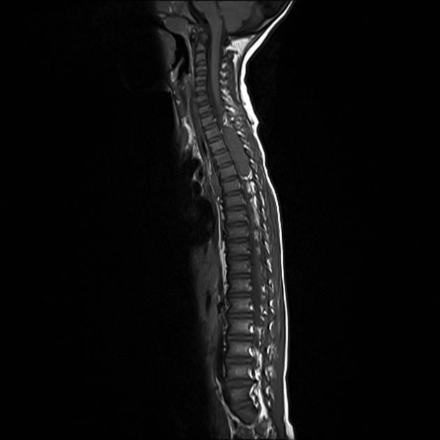
 Subependymal giant cell astrocytoma MRI 3.jpg Sujit Routray
00:24, 3 November 2015
387 × 512; 121 KB
-
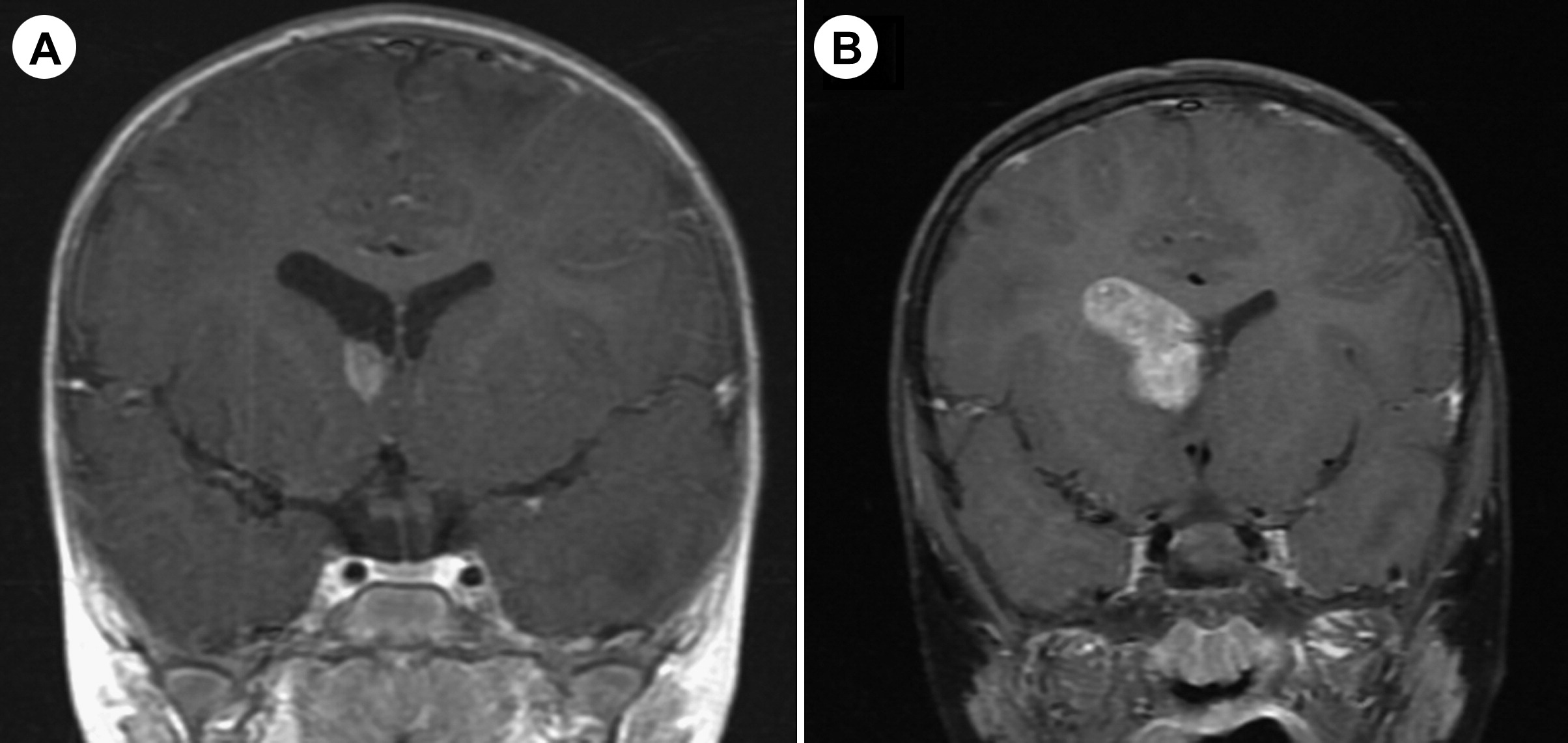 Subependymal giant cell astrocytoma MRI 2.jpg Sujit Routray
00:08, 3 November 2015
2,408 × 1,141; 288 KB
-
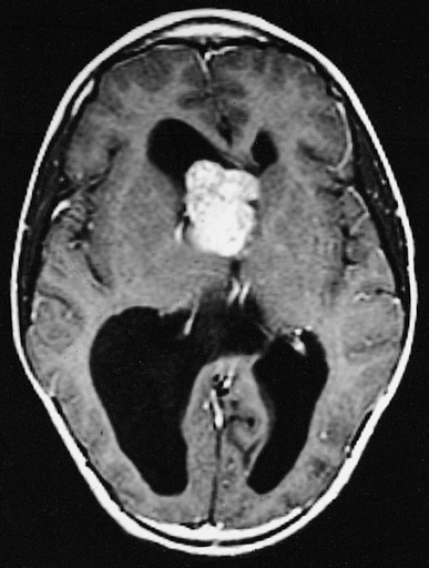
 Subependymal giant cell astrocytoma MRI 1.jpg Sujit Routray
00:02, 3 November 2015
2,408 × 1,074; 291 KB
-
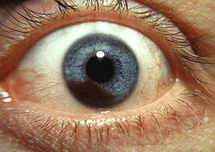
 Proptosis and lid retraction from Graves' Disease.jpg Jyostna Chouturi
19:18, 2 November 2015
300 × 198; 50 KB
-

-
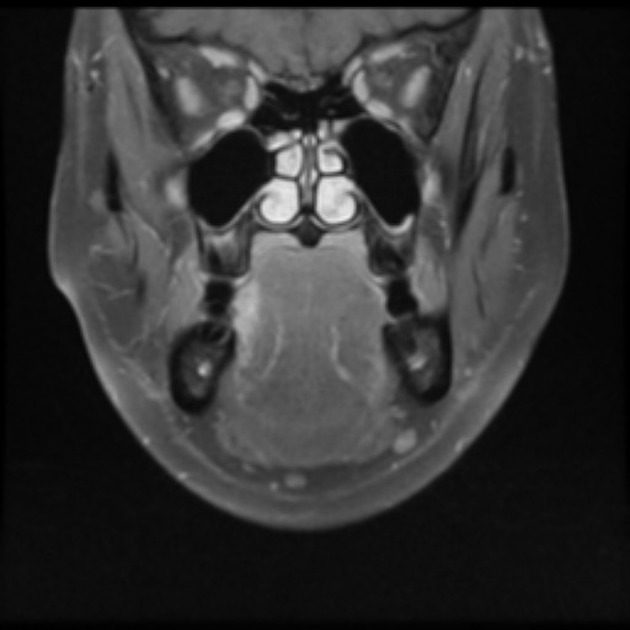
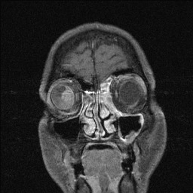
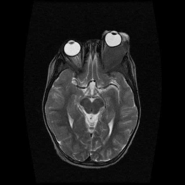
 Optic glioma showing massive proptosis on MRI.jpg Sujit Routray
23:41, 30 October 2015
442 × 512; 141 KB
-
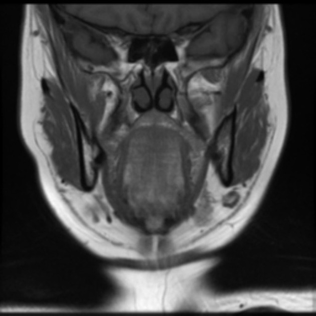
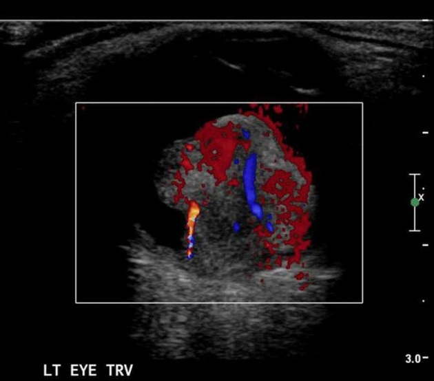
 Proptosis in pilocytic astrocytoma in optic nerve tumor.jpg Sujit Routray
23:35, 30 October 2015
686 × 512; 220 KB
-
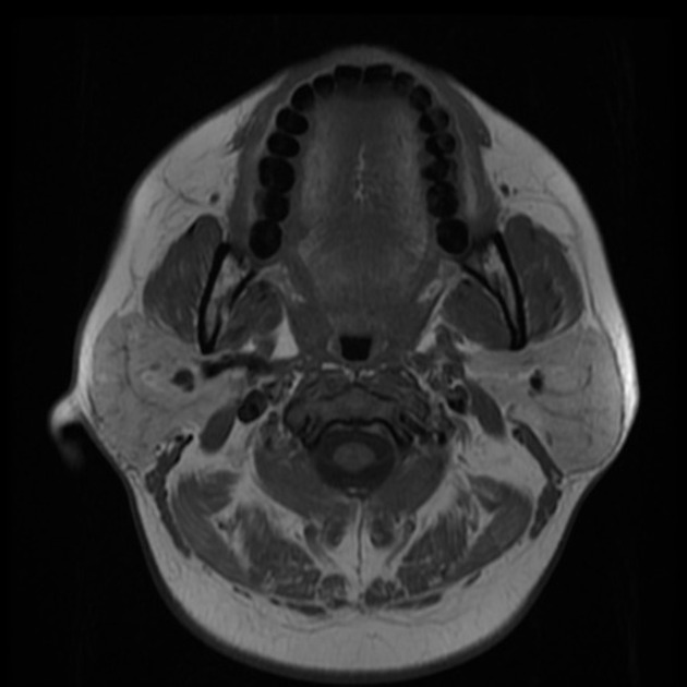
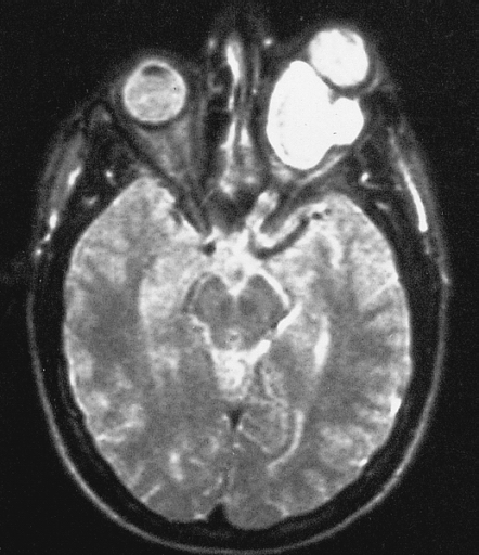
 Pilocytic astrocytoma of the hypothalamic region.jpg Sujit Routray
23:26, 30 October 2015
562 × 512; 190 KB
-
 Genetic mutations in pilocytic astrocytoma.jpg Sujit Routray
19:00, 30 October 2015
778 × 954; 330 KB
-
-
-
-
-

-

-
 800px-Hairy cell leukemia - very high mag.jpg Haytham Allaham
18:48, 29 October 2015
800 × 533; 140 KB
-
-
-
-
-
-
-
-
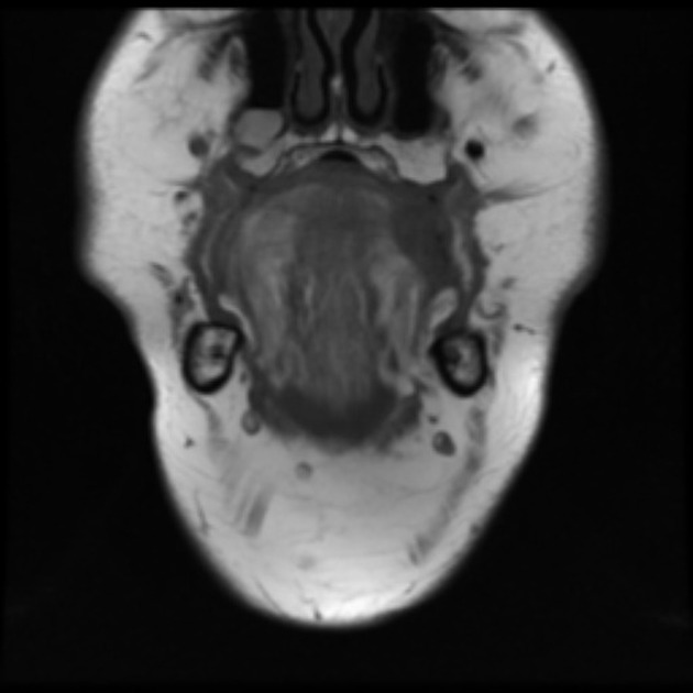
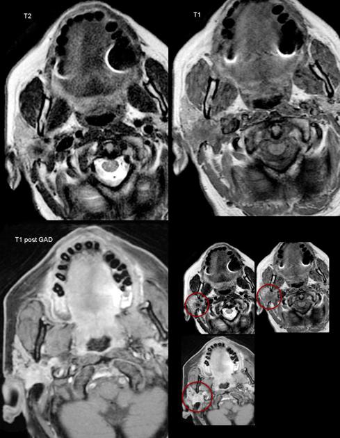
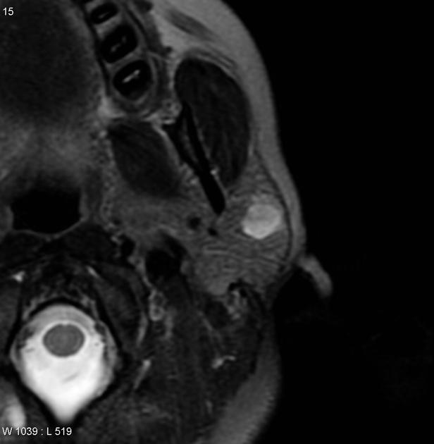
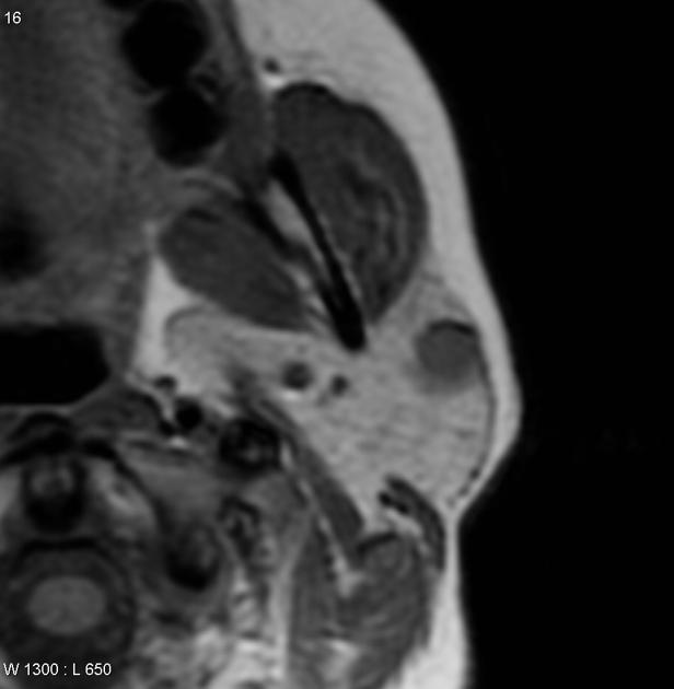
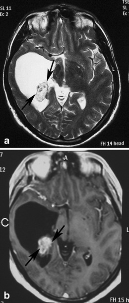
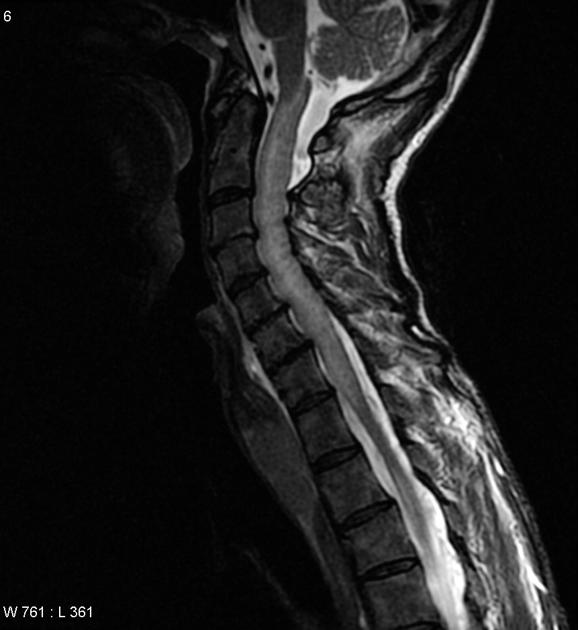
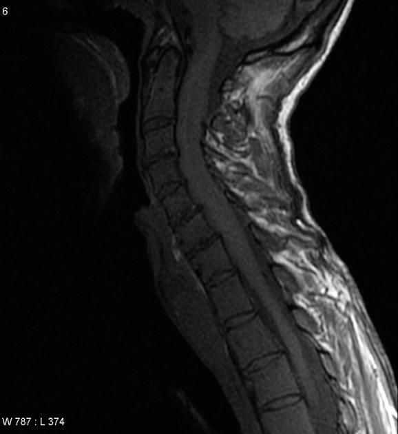
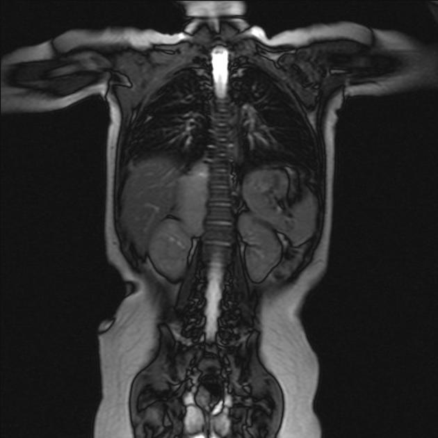
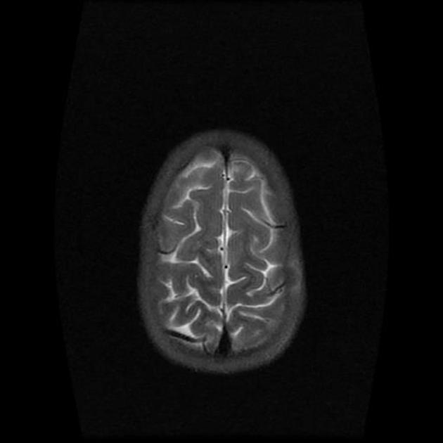
 MRI Follicular thyroid cancer brain metastasis.jpg Ammu Susheela
20:11, 28 October 2015
587 × 630; 45 KB
-

-
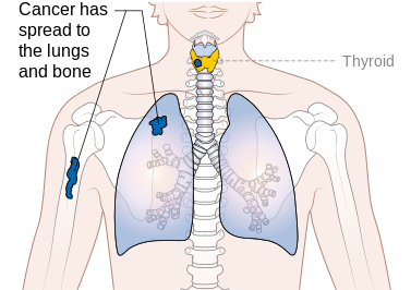
 Follicular thyroid cancer lung metastasis.jpeg Ammu Susheela
20:00, 28 October 2015
630 × 504; 52 KB
-

-
 Cervical metastasis of follicular thyroid cancer.png Ammu Susheela
17:52, 28 October 2015
325 × 235; 65 KB
-
-
-
 Metastatic follicular carcinoma in the bone.png Ammu Susheela
17:26, 28 October 2015
338 × 245; 215 KB
-
 FNAC lymph node metastatic follicular carcinoma 03.png Ammu Susheela
17:21, 28 October 2015
337 × 243; 177 KB
-
 Lymphnode FNAC metastatic follicular carcinoma 02.png Ammu Susheela
17:17, 28 October 2015
342 × 258; 203 KB
-
-
 Lymph node FNAC metastatic follicular carcinoma.png Ammu Susheela
14:22, 28 October 2015
344 × 257; 225 KB
-
-
-
-
 Pilocytic astrocytoma CT scan with hemorrhage.jpeg Sujit Routray
23:13, 27 October 2015
539 × 630; 62 KB
-
 Trans-glottic-squamous-cell-carcinoma-1.jpg Faizan Sheraz
19:23, 27 October 2015
1,024 × 1,024; 57 KB
-
-
-
 Pilocytic astrocytoma endothelial proliferations.jpg Sujit Routray
18:29, 27 October 2015
2,080 × 1,542; 940 KB
-
-

-

-

-

-
-
-
-
-
-

-
-
 Immunohistochemistry of pilocytic astrocytoma Ki-67.jpeg Sujit Routray
00:14, 27 October 2015
998 × 789; 234 KB
-
-
-
-
-
-
-
 Coronal C delayed orbital malignant melanoma.png Simrat Sarai
18:26, 26 October 2015
630 × 589; 180 KB
-
 Axial C delayed choroidal malignant melanoma.png Simrat Sarai
18:22, 26 October 2015
630 × 589; 151 KB
-
 Orbital malignant melanoma Axial C+ delayed.jpg Simrat Sarai
18:12, 26 October 2015
630 × 589; 31 KB
-
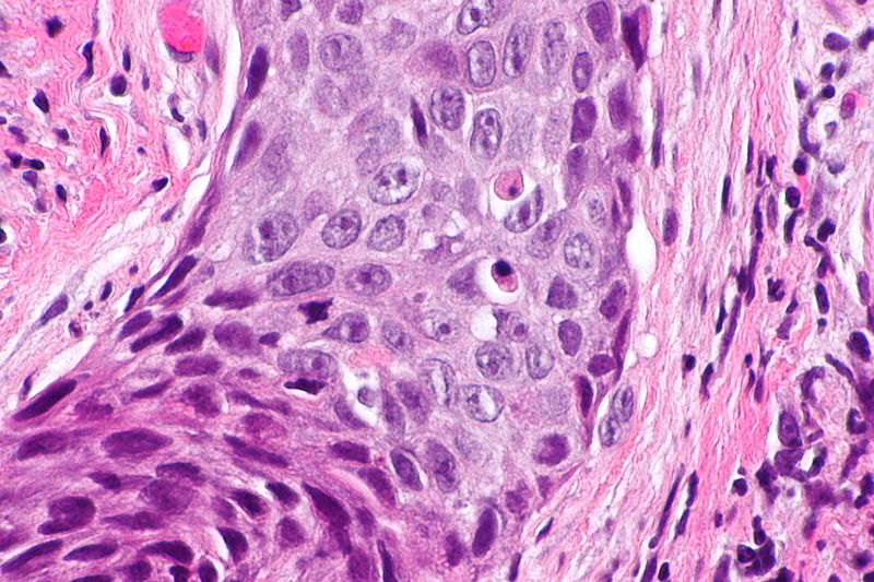 800px-Laryngeal squamous carcinoma -- very high mag.jpg Faizan Sheraz
17:59, 26 October 2015
800 × 533; 129 KB
-
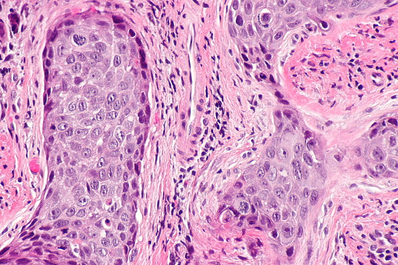 800px-Laryngeal squamous carcinoma -- high mag.jpg Faizan Sheraz
17:55, 26 October 2015
800 × 533; 151 KB
-
-
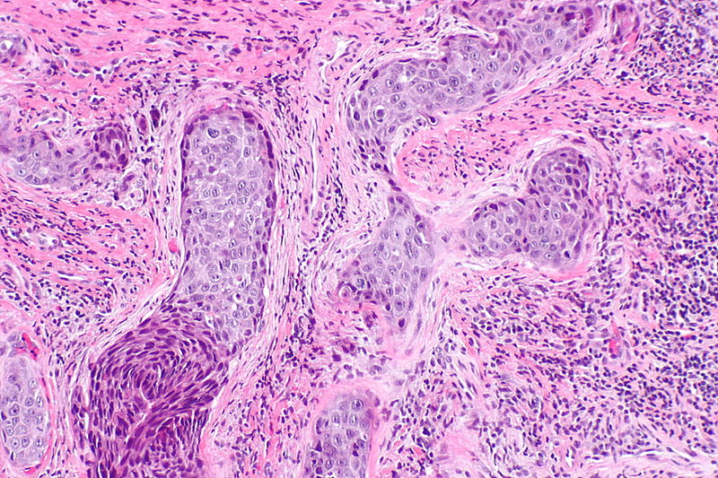 800px-Laryngeal squamous carcinoma -- intermed mag.jpg Faizan Sheraz
17:50, 26 October 2015
800 × 533; 183 KB
-
 Axial C+ delayed orbital malignant melanoma.jpg Simrat Sarai
20:28, 23 October 2015
630 × 589; 31 KB
-
 Coronal C+ delayed orbital malignant melanoma.jpg Simrat Sarai
20:21, 23 October 2015
630 × 589; 35 KB
-

-
-

-

-
-

-

-
-

-

-

-
-
-

-
-
-

-

-
-

-

-
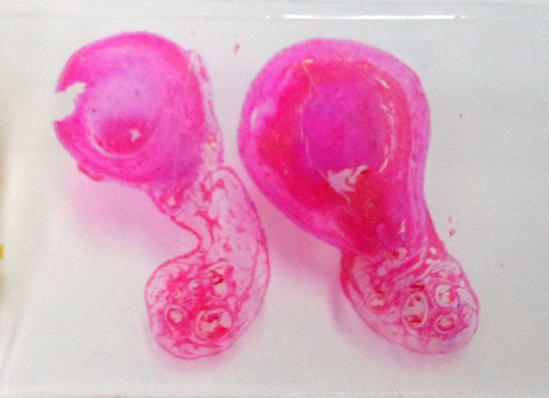
 193px-Hydatidiform mole (1) complete type.jpg Monalisa Dmello
14:42, 20 October 2015
193 × 145; 17 KB
-
-

-
-
-
-
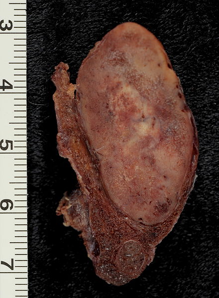
 Metastatic follicular thyroid carcinoma - Case 264 (8558730243).jpg Ammu Susheela
22:50, 19 October 2015
800 × 600; 115 KB
-
 Metastatic follicular thyroid carcinoma - Case 264.jpg Ammu Susheela
22:34, 19 October 2015
800 × 600; 102 KB
-
 Papillary thyroid carcinoma oncocytic variant -- very high mag.jpg Ammu Susheela
22:32, 19 October 2015
800 × 533; 116 KB
-
 Papillary thyroid carcinoma oncocytic variant -- high mag.jpg Ammu Susheela
22:29, 19 October 2015
800 × 533; 131 KB
-
-
 Thyroid PapillaryCarcinoma CribriformMorularVariant04.jpg Ammu Susheela
22:10, 19 October 2015
800 × 600; 75 KB
-

-
 St. Louis Encephalitis (SLE) virus EM PHIL 1871 lores.JPG Turky Alkathery
19:53, 19 October 2015
1,024 × 1,331; 499 KB
-
-
-
-
 Papillary thyroid cancer orphan annie eye nucleus.jpg Ammu Susheela
22:08, 18 October 2015
630 × 502; 64 KB
-
-

-
-
-
-
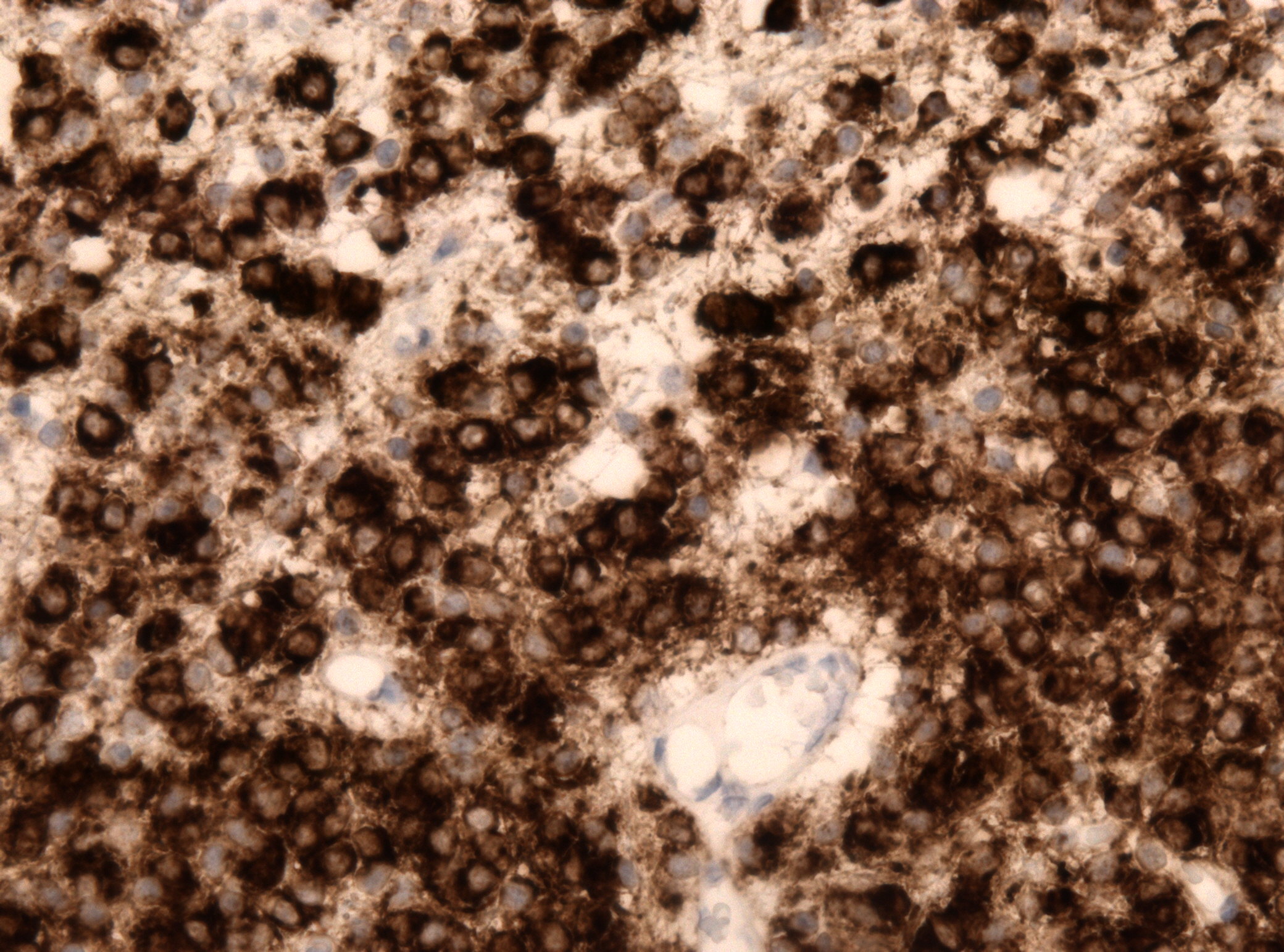
 800px-Appendix Carcinoid Synaptophysin 14BR---.jpg Faizan Sheraz
15:49, 16 October 2015
800 × 600; 86 KB
-
-
-
-

-

-

-
-
-
-
-

-

-
-
-

-
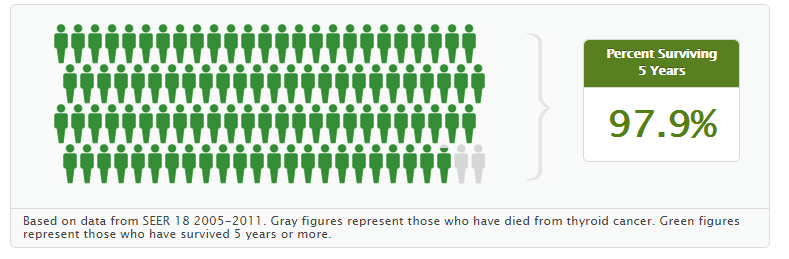
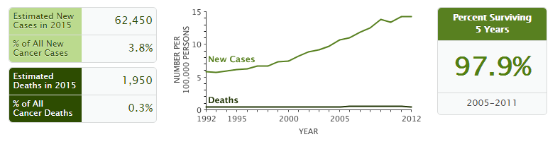
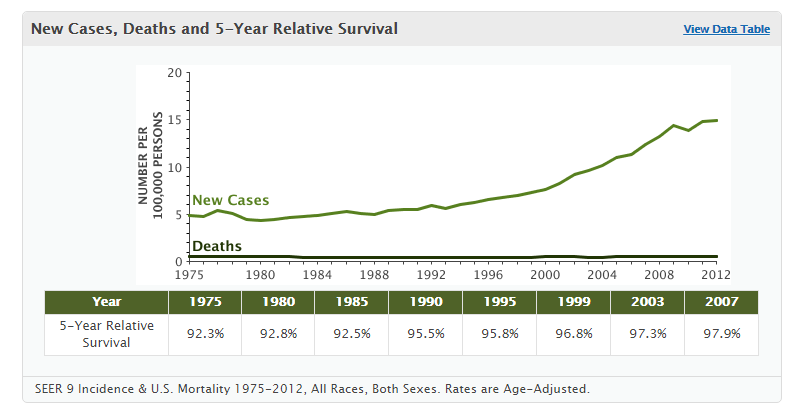 New cases, deaths and 5 year relative survival.PNG Ammu Susheela
20:41, 14 October 2015
799 × 414; 27 KB
-
-

-
-
-
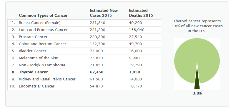
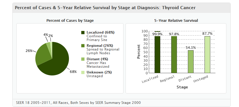 5 year survival pie chart for thyroid cancer.PNG Ammu Susheela
20:06, 14 October 2015
832 × 373; 37 KB
-
-
-
-
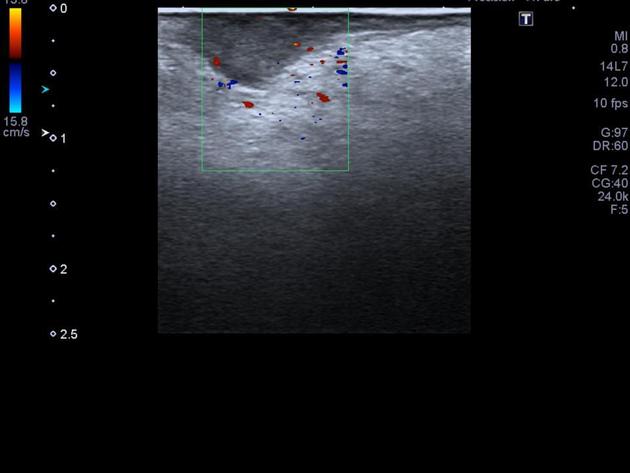
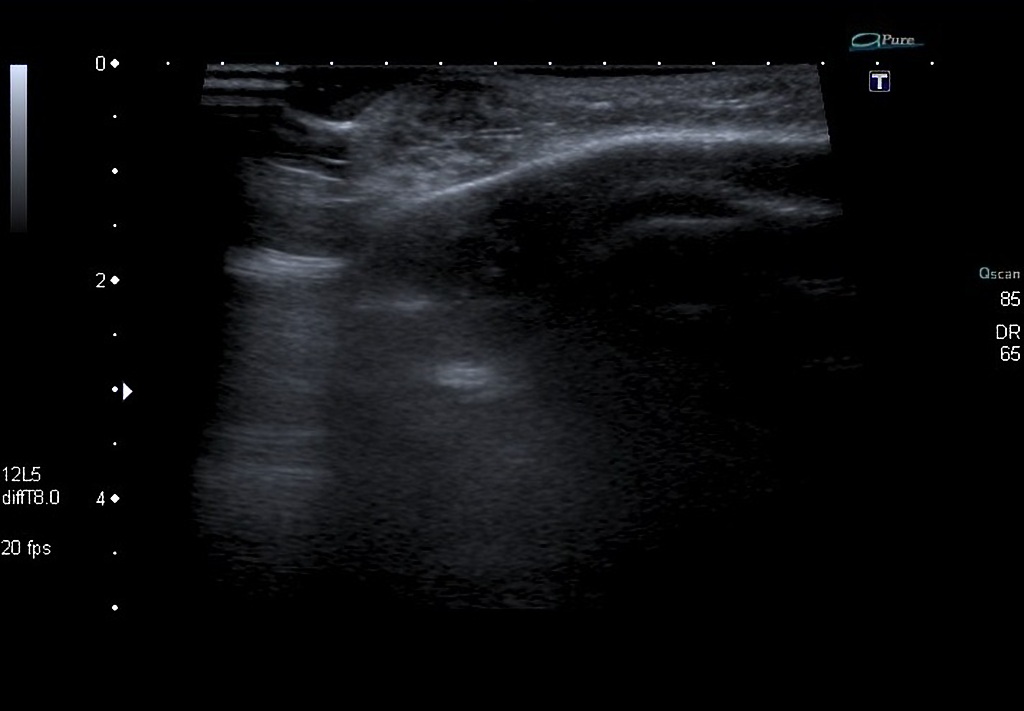
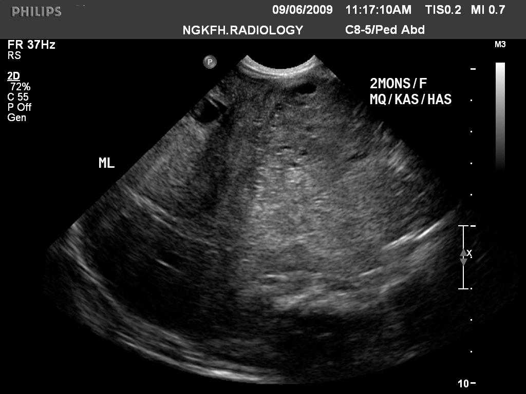
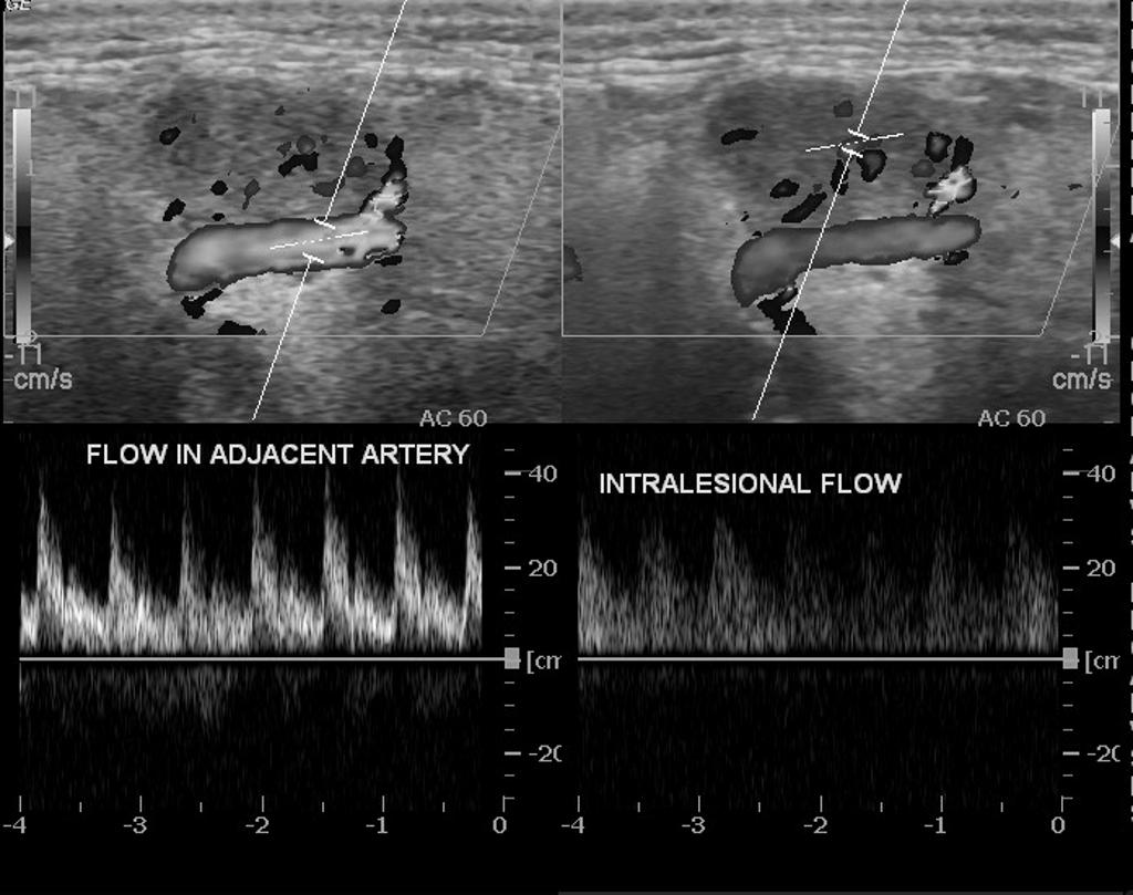
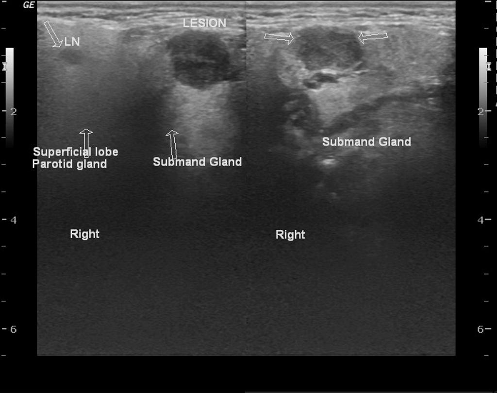
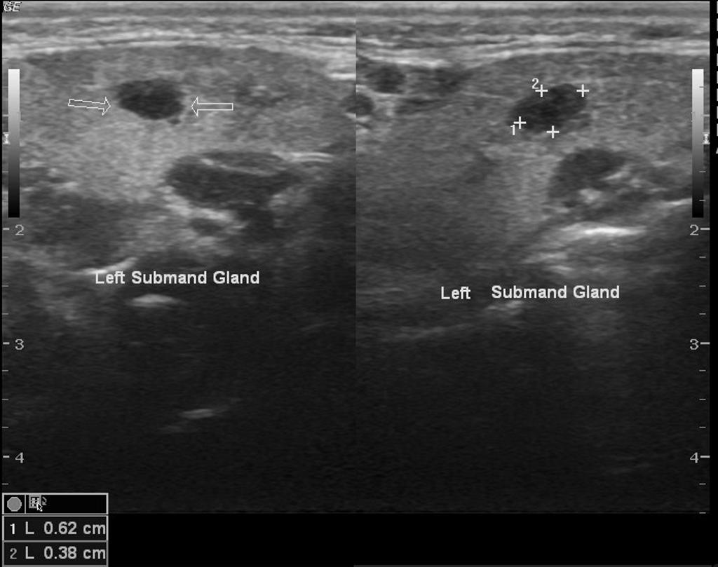
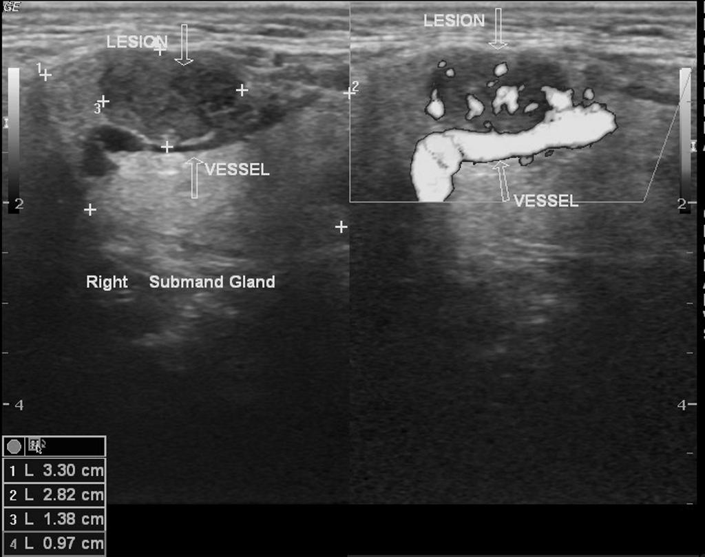
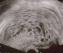
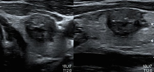
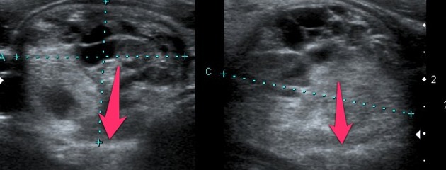
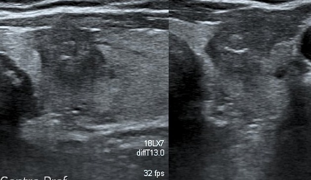
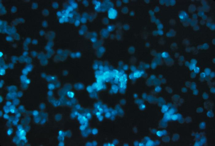
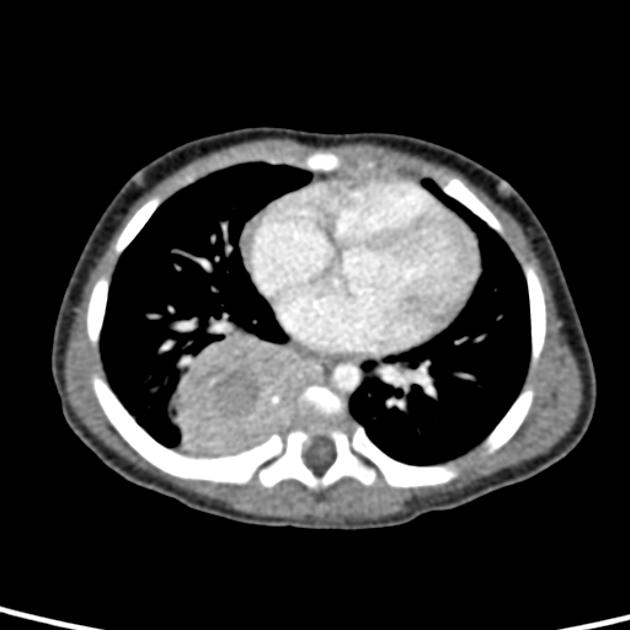
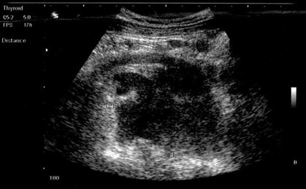 Anaplastic large cell lymphoma Ultrasound.JPG Sowminya Arikapudi
19:19, 14 October 2015
630 × 389; 40 KB
-
-
-
-
-
-

-
-
-
-
-
-
-
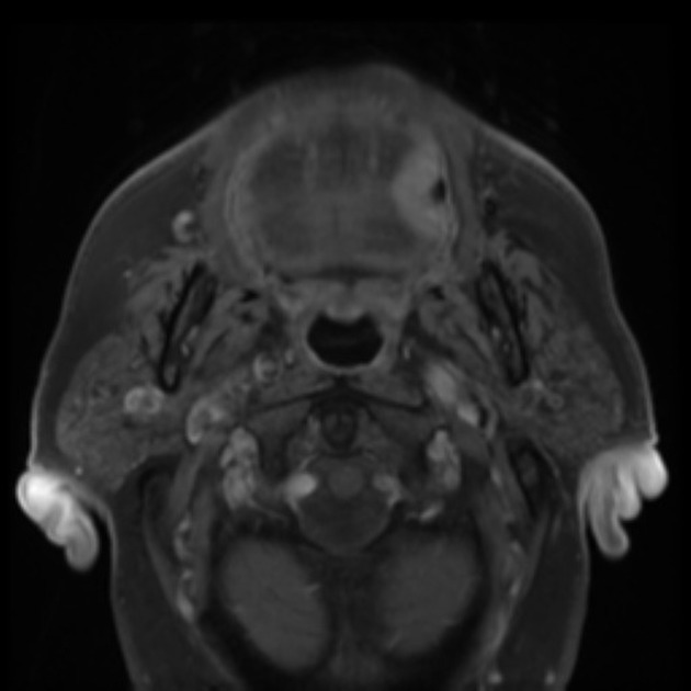
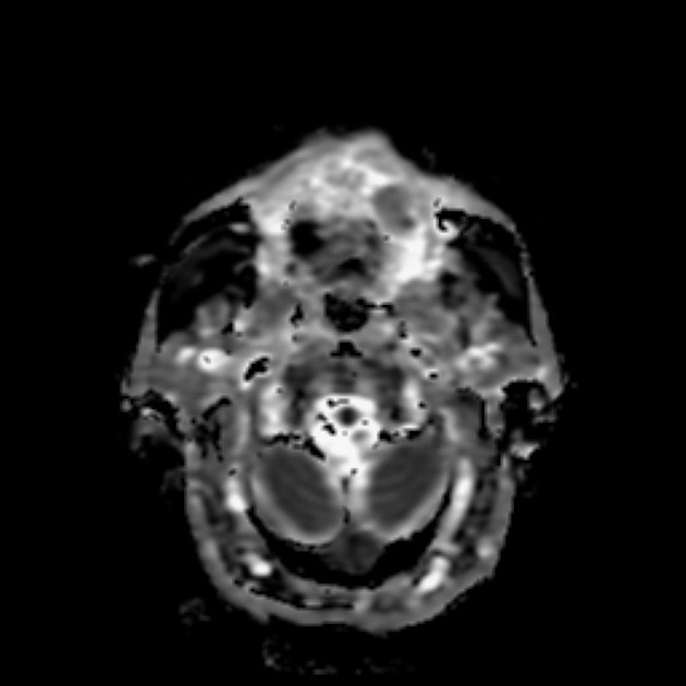
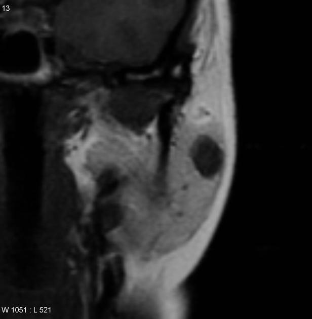
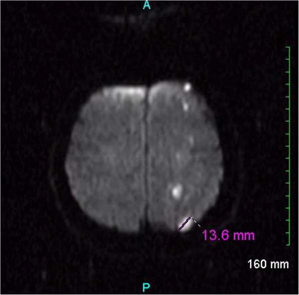
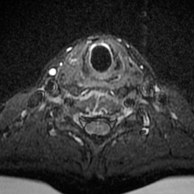
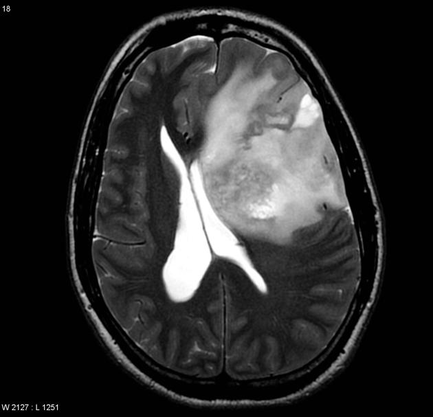
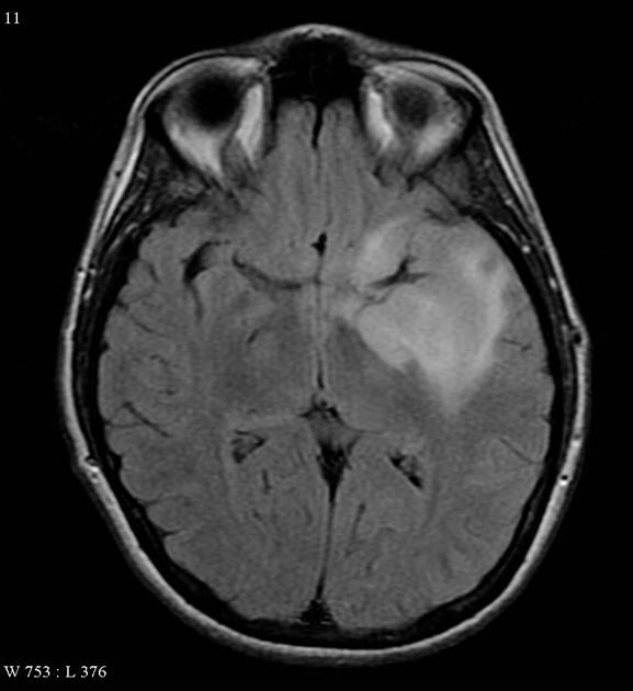
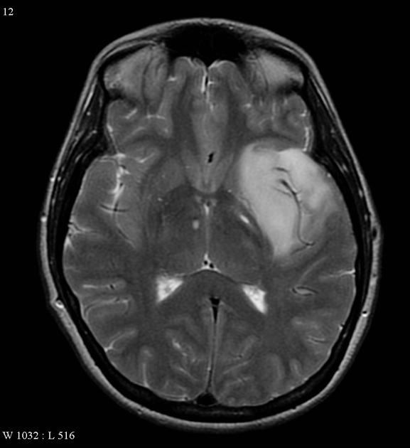
 Anaplastic large cell lymphoma MRI scan .png Sowminya Arikapudi
14:46, 14 October 2015
630 × 364; 87 KB
-
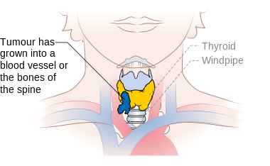 Diagram showing stage T4b thyroid cancer CRUK 273.png Ammu Susheela
14:40, 14 October 2015
380 × 234; 41 KB
-
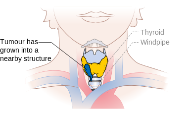 Diagram showing stage T4a thyroid cancer CRUK 272.png Ammu Susheela
14:37, 14 October 2015
380 × 234; 39 KB
-
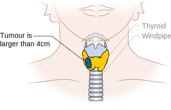 Diagram showing stage T3 thyroid cancer CRUK 265.png Ammu Susheela
14:34, 14 October 2015
342 × 221; 29 KB
-
 Diagram showing stage T2 thyroid cancer CRUK 258.png Ammu Susheela
14:31, 14 October 2015
342 × 221; 29 KB
-
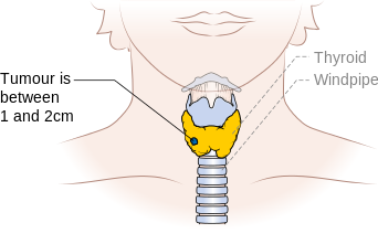 Diagram showing stage T1b thyroid cancer CRUK 251.svg.png Ammu Susheela
14:20, 14 October 2015
342 × 221; 29 KB
-
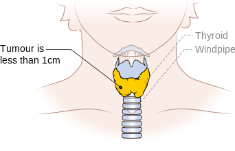 Diagram showing stage T1a thyroid cancer CRUK 250.png Ammu Susheela
14:11, 14 October 2015
342 × 221; 29 KB
-
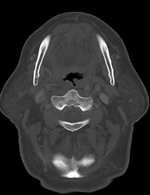
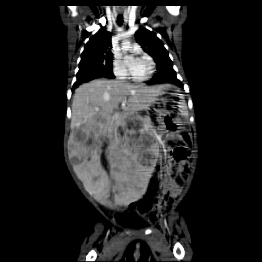
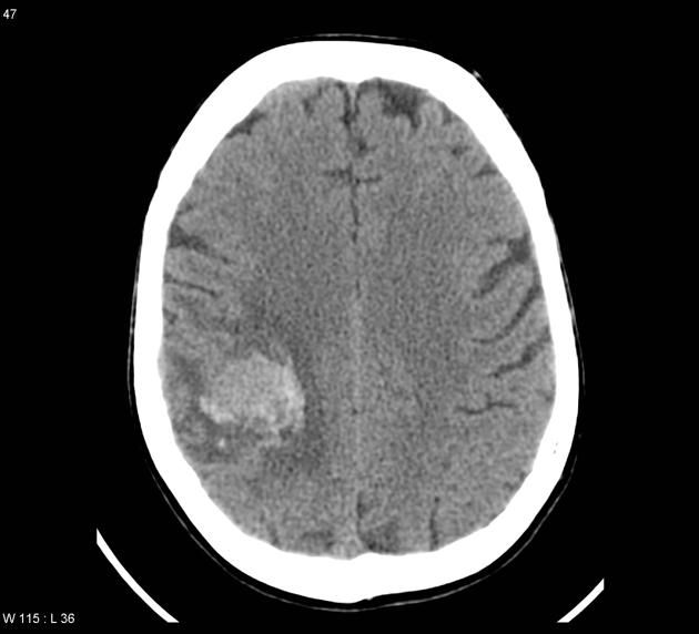
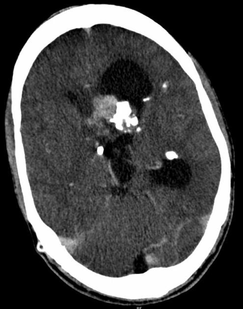
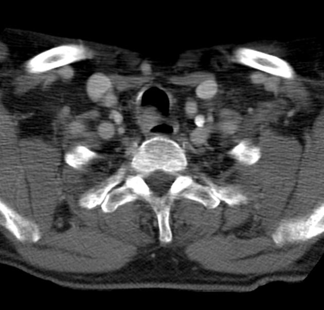
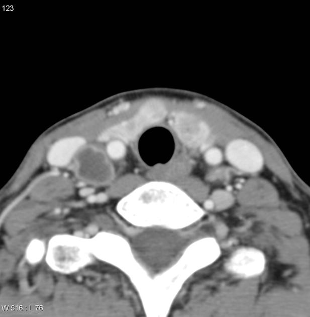
 Anaplastic large cell lymphoma CT scan .png Sowminya Arikapudi
14:04, 14 October 2015
630 × 425; 104 KB
-
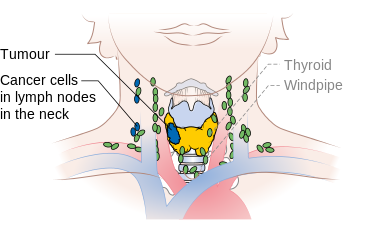 Diagram showing stage N1b thyroid cancer CRUK 243.png Ammu Susheela
14:00, 14 October 2015
390 × 234; 47 KB
-
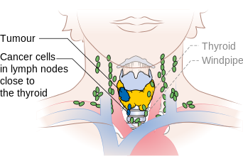 Diagram showing stage N1a thyroid cancer CRUK 242.png Ammu Susheela
13:55, 14 October 2015
342 × 234; 47 KB
-
-

-
-
-

-

-
-
-
-
 Chronic lymphocytic leukemia compared to all cancers in the US.png Haytham Allaham
22:03, 13 October 2015
761 × 436; 44 KB
-
 Chronic lymphocytic leukemia incidence and mortality 1975-2011.png Haytham Allaham
22:02, 13 October 2015
761 × 391; 38 KB
-
 Chronic lymphocytic leukemia incidence rate.png Haytham Allaham
22:00, 13 October 2015
760 × 467; 44 KB
-
 Chronic lymphocytic leukemia incidence rate per age group.png Haytham Allaham
21:59, 13 October 2015
759 × 398; 42 KB
-
 Chronic lymphocytic leukemia mortality rate.png Haytham Allaham
21:57, 13 October 2015
762 × 449; 44 KB
-
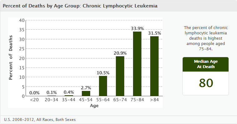 Chronic lymphocytic leukemia mortality rate per age group.png Haytham Allaham
21:55, 13 October 2015
759 × 397; 42 KB
-
 Chronic lymphocytic leukemia survival rate.png Haytham Allaham
21:48, 13 October 2015
760 × 247; 26 KB
-

-

-

-

-
-
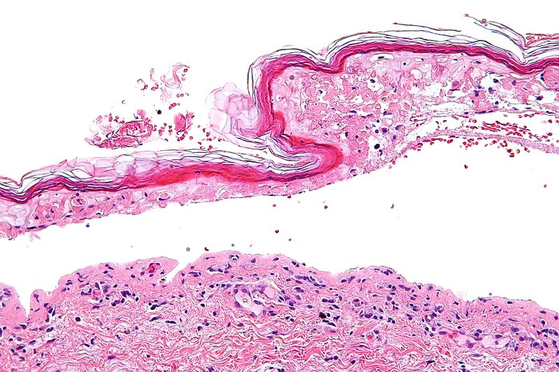
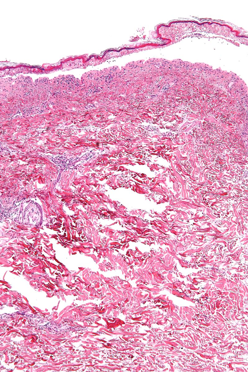 800px-Confluent epidermal necrosis - low mag.jpg Parminder Dhingra
19:34, 13 October 2015
800 × 1,200; 328 KB
-
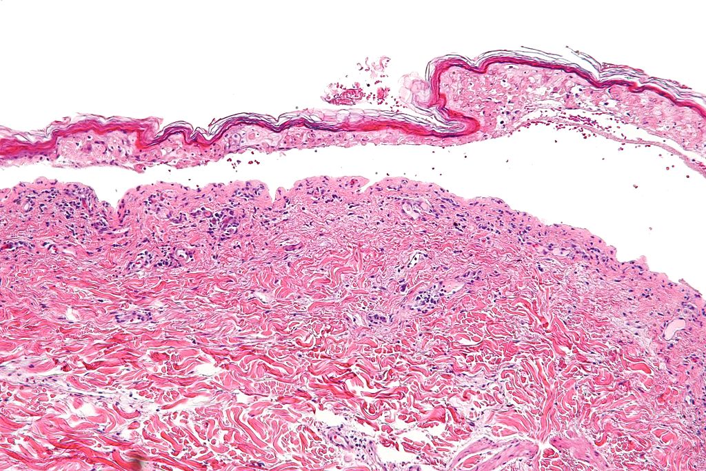 1024px-Confluent epidermal necrosis - intermed mag.jpg Parminder Dhingra
19:33, 13 October 2015
1,024 × 683; 224 KB
-
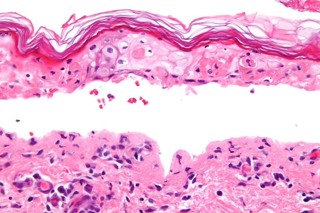 1024px-Confluent epidermal necrosis - very high mag.jpg Parminder Dhingra
19:31, 13 October 2015
1,024 × 683; 121 KB
-
-
 800px-Confluent epidermal necrosis - high mag.jpg Parminder Dhingra
19:29, 13 October 2015
800 × 533; 119 KB
-
 Chronic lymphocytic leukemia - very high mag.jpg Haytham Allaham
17:58, 13 October 2015
800 × 533; 152 KB
-
 Chronic lymphocytic leukemia - high mag.jpg Haytham Allaham
17:54, 13 October 2015
800 × 533; 195 KB
-
 Chronic lymphocytic leukemia - intermed mag.jpg Haytham Allaham
17:51, 13 October 2015
800 × 533; 233 KB
-
-
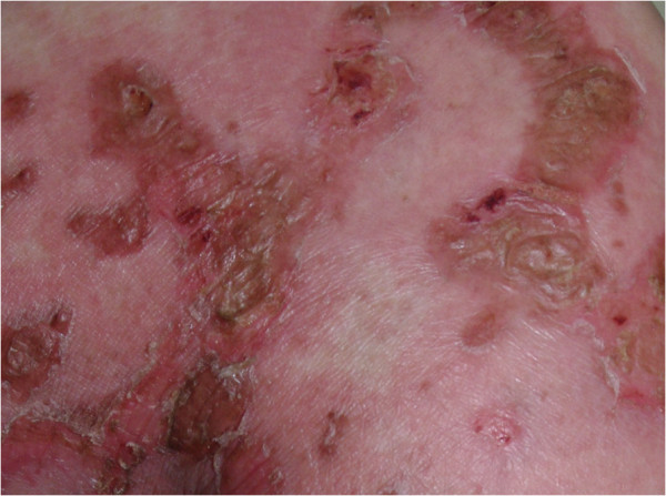 Close-up view of well demarcated erythematous plaques, with fragile vesicles on gluteal area..jpg Sujit Routray
15:28, 13 October 2015
600 × 447; 102 KB
-
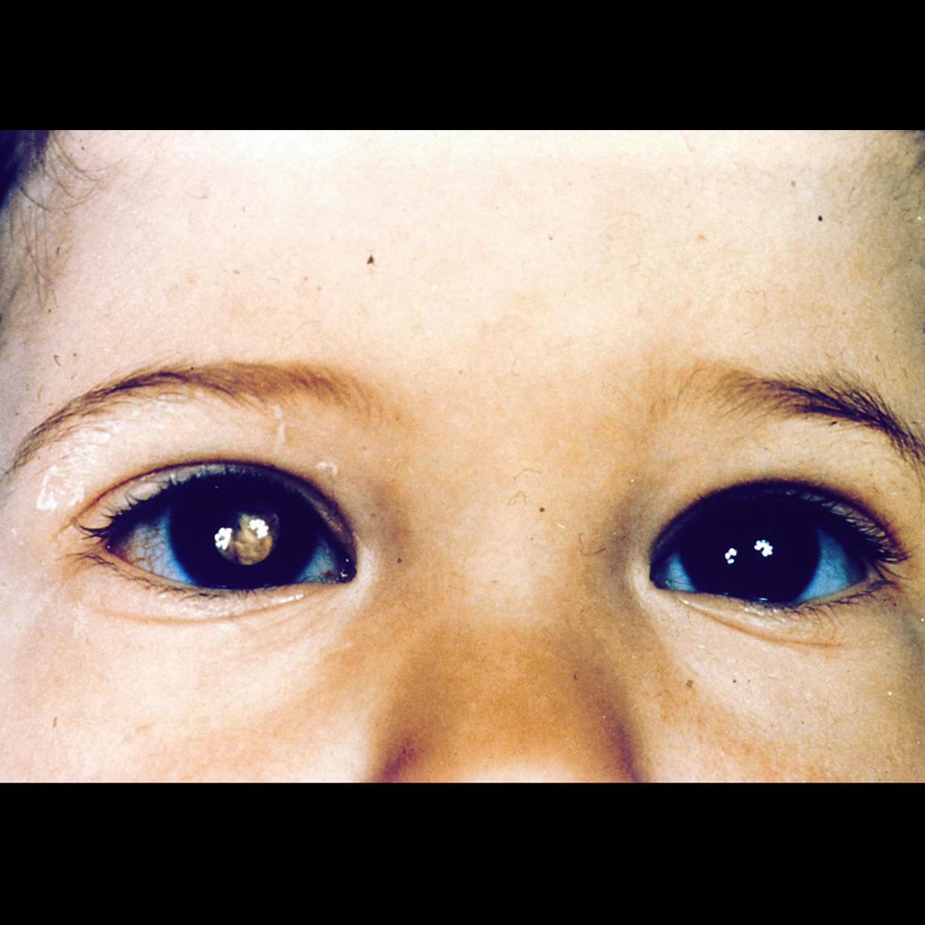
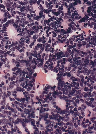 Flexner- Wintersteiner Rosettes in Retinoblastoma.jpg Simrat Sarai
19:03, 12 October 2015
368 × 512; 192 KB
-
-
-
-
-
-

-

-
-
-
-
-
-

-
-
-
-
-
-
-
-
-
-
-
-
-

-

-
-
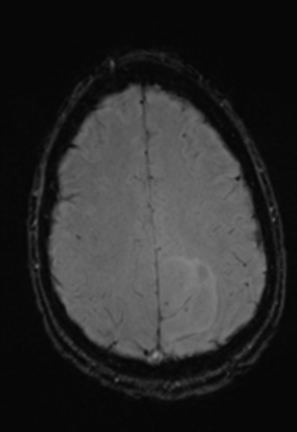
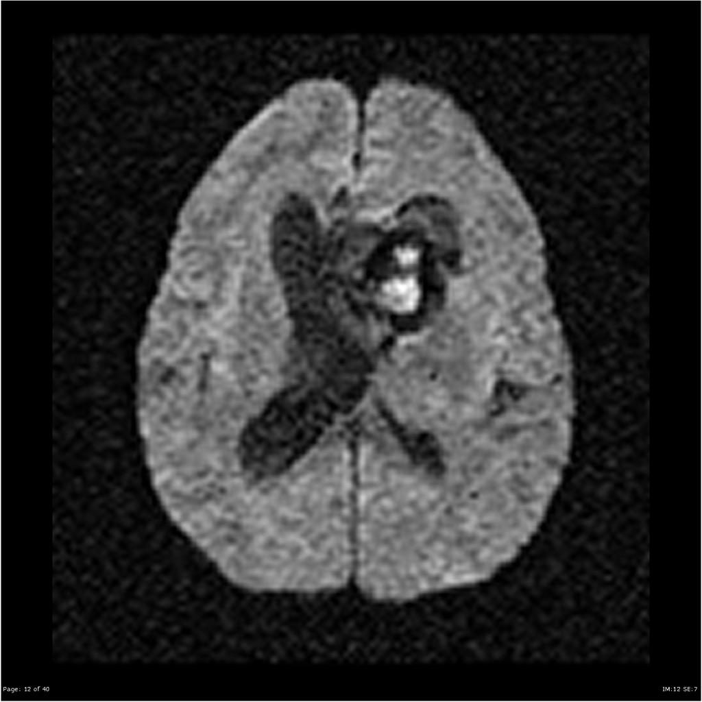
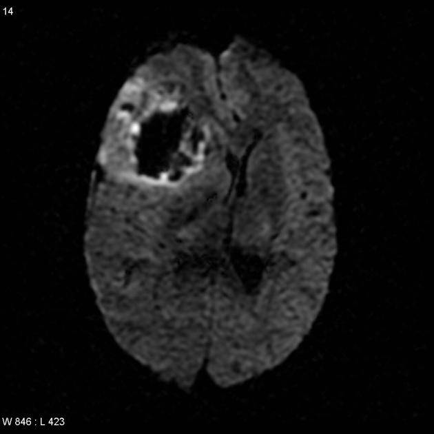 Oligodendroglioma - anaplastic - haemorrhagic MRI axial DWI.jpg Sujit Routray
17:15, 8 October 2015
630 × 630; 22 KB
-

-
-

-

-
-
-
 IDH1 R132H in anaplastic ologodendroglioma.jpg Sujit Routray
14:27, 8 October 2015
2,080 × 1,542; 697 KB
-

-
 Oligodendroglioma discrete invasion HE.jpg Sujit Routray
14:15, 8 October 2015
2,080 × 1,542; 1.11 MB
-

-
 Anaplastic oligodendroglioma minigemistocytes.jpg Sujit Routray
13:21, 8 October 2015
800 × 593; 106 KB
-

-
-
-
-

-

-
-
-
 73c54460c3e8d80f3d18cd522ed152 big gallery.jpg Ahmad Al Maradni
17:50, 7 October 2015
630 × 630; 37 KB
-
 A91344c612b07b087964a93ee047df big gallery.jpg Ahmad Al Maradni
16:42, 7 October 2015
630 × 630; 42 KB
-
-
-

-

-
-

-
 Medulloblastoma nuclear moulding HE stain.jpg Haytham Allaham
18:30, 6 October 2015
800 × 593; 158 KB
-

-
-
-
 Medulloblastoma stratum moleculare x40 (1).jpg Haytham Allaham
18:19, 6 October 2015
800 × 593; 187 KB
-
 Desmoplastic medulloblastom MIB1 proliferation.jpg Haytham Allaham
18:15, 6 October 2015
800 × 593; 152 KB
-

-
 Desmoplastic medulloblastoma reticulin stain pale island.jpg Haytham Allaham
18:12, 6 October 2015
800 × 593; 185 KB
-
-
-
-
 Ependymoma-true-ependymal-rosettes123.jpg Ahmad Al Maradni
16:08, 6 October 2015
1,024 × 1,024; 95 KB
-
-
-
 120px-Adrenal Neuroblastoma VascularInvasion MP CTR.jpg Haytham Allaham
01:50, 6 October 2015
120 × 90; 6 KB
-
-
-
-
-
-
-


.jpeg)


.jpg)
.jpg)


_in_adult.jpg)


_parotid_gland.jpg)
_HE_stain_(1).jpg)

.jpg)


.jpg)
.jpg)
.jpg)
.jpg)


.jpg)


.jpg)


_complete_type.jpg)


.jpg)


_virus_EM_PHIL_1871_lores.JPG)


.jpg)


.jpg)


.jpg)
.jpg)

.jpg)
.jpg)


.jpg)


.jpg)


_in_adult.JPG)

_in_adult.JPG)


