Sick sinus syndrome EKG examples: Difference between revisions
No edit summary |
|||
| Line 4: | Line 4: | ||
'''For the main page on sick sinus syndrome, click [[Sick sinus syndrome|here]].''' | '''For the main page on sick sinus syndrome, click [[Sick sinus syndrome|here]].''' | ||
==EKG examples== | ==EKG examples== | ||
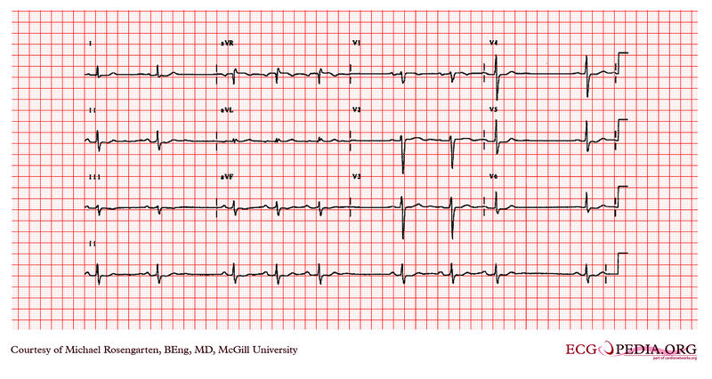
Shown below is an EKG recorded in a middle-aged woman treated for [[atrial fibrillation]]. The patient was doing well when this [[electrocardiogram]] was taken, and was taking [[digoxin]] and [[flecainide]] . The [[electrocardiogram]] shows an irregular rhythm which appears to be sinus. The grouping of the [[QRS]] complexes suggests a [[Mobitz type I AV block]]. In this case though, no [[AV block]] is seen and this may represent a | Shown below is an EKG recorded in a middle-aged woman treated for [[atrial fibrillation]]. The patient was doing well when this [[electrocardiogram]] was taken, and was taking [[digoxin]] and [[flecainide]] . The [[electrocardiogram]] shows an irregular rhythm which appears to be sinus. The grouping of the [[QRS]] complexes suggests a [[Mobitz type I AV block]]. In this case though, no [[AV block]] is seen and this may represent a [[sinus node exit block]]. | ||
[[Image:Sick sinus syndrome2.jpg|center|800px]] | [[Image:Sick sinus syndrome2.jpg|center|800px]] | ||
Copyleft image obtained courtesy of ECGpedia, http://en.ecgpedia.org/wiki/Main_Page | Copyleft image obtained courtesy of ECGpedia, http://en.ecgpedia.org/wiki/Main_Page | ||
Latest revision as of 23:12, 24 October 2012
Editor-In-Chief: C. Michael Gibson, M.S., M.D. [1]
For the main page on sick sinus syndrome, click here.
EKG examples
Shown below is an EKG recorded in a middle-aged woman treated for atrial fibrillation. The patient was doing well when this electrocardiogram was taken, and was taking digoxin and flecainide . The electrocardiogram shows an irregular rhythm which appears to be sinus. The grouping of the QRS complexes suggests a Mobitz type I AV block. In this case though, no AV block is seen and this may represent a sinus node exit block.

Copyleft image obtained courtesy of ECGpedia, http://en.ecgpedia.org/wiki/Main_Page
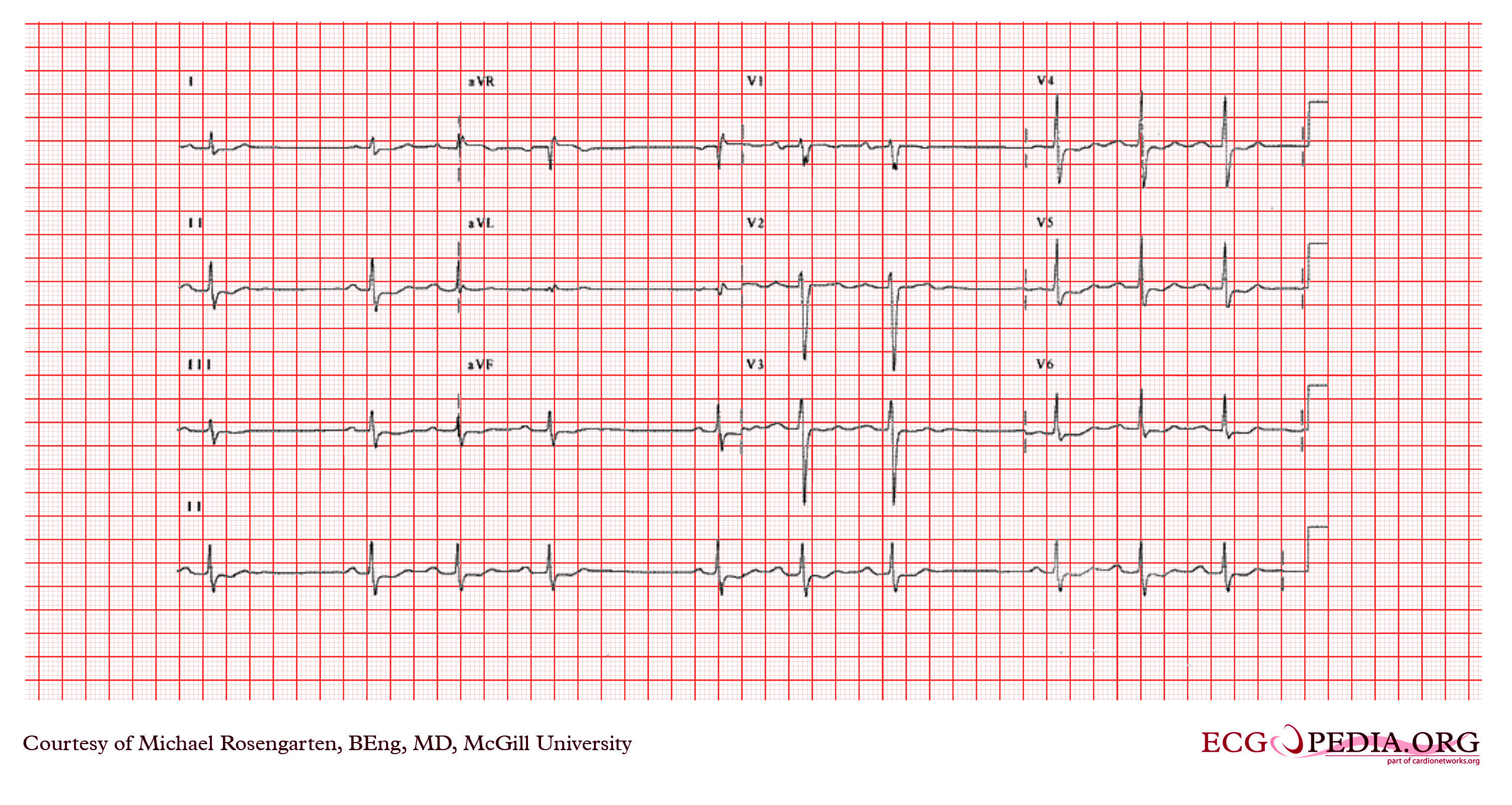
Shown below is an EKG recording from a middle aged woman with recurrent atrial fibrillation. She was treated with flecainide with some improvement of her symptoms. The above is a recording taken in the cardiac clinic. It shows group beating with sinus rhythm. There are missing P waves. This suggests that there is sinus node arrest or that this is sinus node exit block.

Copyleft image obtained courtesy of ECGpedia, http://en.ecgpedia.org/wiki/Main_Page
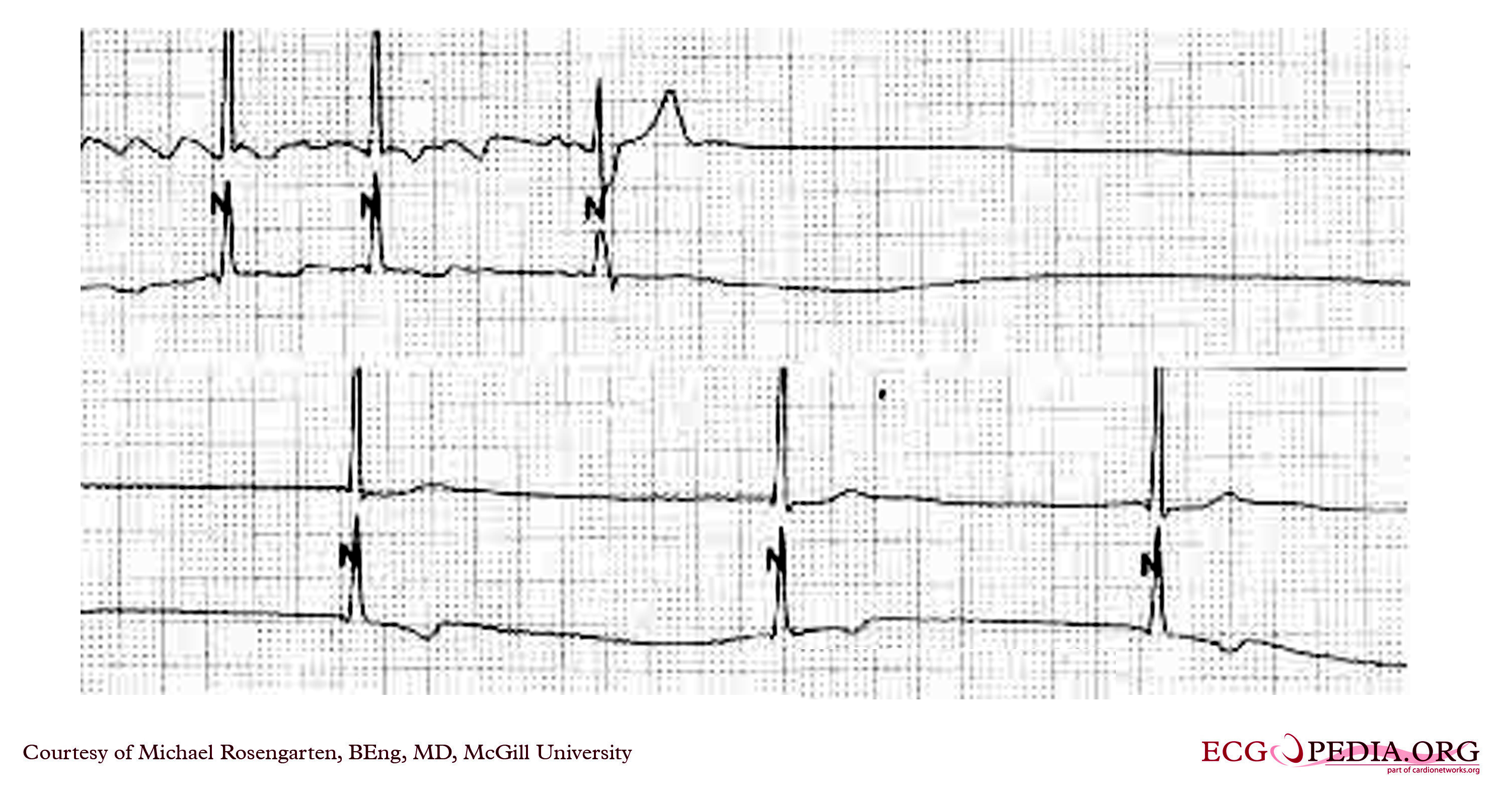
Shown below is a Holter recording from a patient that illustrates a bradycardia after the termination of a tachycardia. This has been called the "tachy/brady syndrome". In this case the tachy part is atrial fibrillation, which is followed by a long pause and then a nodal escape at about 35/min. Patients are often symptomatic from the pause and may have dizziness or syncope on termination of the tachycardia.

Copyleft image obtained courtesy of ECGpedia, http://en.ecgpedia.org/wiki/Main_Page