Sandbox: Reddy
Overview
Historical Perspective
- In 1880, Herman and Desfosses described the anal glands within the internal sphincter, sub-mucosa and their opening into the anal crypts and demonstrated that the infection of these glands and the spread of the infection through the intersphincteric space can result in the formation of a anal abscess.[1]
- Tucker and Hellwig, provided evidence that the initial infection occurs in the anal ducts allowing the infection to spread from the anal lumen into the anal canal wall.[1]
- In 1950, Goligher described the treatment for anal abscess with inicision and curettage with antibiotic bath and primary closure.[1]
Classification
Based on the location of the abscess in relation to the anal canal and the spread of infection to the surrounding structures anal abscess can be classified into [2]
- Perianal abscess:When the infection reaches the anal verge passing between the internal and external sphincter, it results in the formation of a perianal abscess.
- Ischiorectal abscess:If the infection ruptures through the external sphincter it results in a formation of a ischiorectal abscess.
- Supralevator abscess:If the infection extends superiorly, it can form a supralevator abscess.
- Horseshoe abscess:Extension of the anal abscess to both the ischiorectal fossa results in the formation of a horseshoe abscess.
Based on the location the abscesses can also be classified into:
- High anorectal abscess: These include intersphincteric, perianal, and ischiorectal abscesses.
- Low anorectal abscess: These incude submucosal, supralevator abscesses.
Pathophysiology
Pathogenesis
- Anal canal is a 2 to 4cm in length, starts at the anorectal junction to the end of anal verge.[3]
- It is divided into a upper and a lower part by transition zone that is seen at the dentate or pectinate line which is surrounded by longitudinal mucosal folds, called columns of morgagni.[3]
- Each of this fold contains anal crypts, each of which contains 3 to 12 anal glands, the distribution of these glands is not uniform with most of the glands present anterior to the position of the anal canal and fewer in the posterior position.[3]
- The initial infection occurs in the ducts of the anal glands and the spread of infection results in the formation of the abscess, various theories were put forward to describe the pathogenesis and the most accepted one is the cryptoglandular theory.[4]
- The crytoglandular theory states that obstruction of anal gland duct results in a infection and due to the presence of these glands deep in relation to the anal canal and sphincter, the infection follows the path of least resistance resulting in a abscess formation at the termination of the gland.[5]
Causes
- Supralevator abscess can be caused by the spread of infection from abdominal infections such as appendicitis, diverticulitis, or gynecologic sepsis.
- Spread of infection of ano-rectal Crohn's disease.
- Trauma to the anal canal
- Cancer of the anal canal or the anal glands
Microbial Causes
Epidemiology and Demographics
Incidence
- The incidence of anorectal abscess is estimated to be around 68,000 to 96,000 cases per year in the United States.[1]
Gender
- Anorectal abscesses are two times more frequently seen in men than women.[6]
Age
- Patients with anal abscess present between ages of 20 to 60 years with a mean age of 40 in both sexes.[1]
Race
- There are limited epidemiological studies which studied the frequency of anal abscess with race differences, however a study in Chicago showed that 92% of the patients presented with anal abscess were African population.[7]
Risk Factors
Risk factors for the development of recurrent of anal abscesses include[8]:
- Crohn's disease
- Diabetes mellitus
- History of abscess in the ischiorectal location
- HIV infection
- Receptive anal sex
Differential Diagnosis
Anorectal abscess must be differentiated from other causes of anal pain including anal fissure, thrombosed hemorrhoids, levator spasm, sexually transmitted disease, proctitis, and hidradenitis suppurativa, infected skin furuncles, herpes simplex virus, HIV, tuberculosis, syphilis,and actinomycosis
- Cancer[9]
Natural History, Prognosis, Complications
Natural History
Complications
- Recurrence usually to incomplete drainage of the abscess
- Sepsis
- Fistula formation
- Scarring
Diagnosis
History and Symptoms
- Patients with low abscess present with usually presents with anal pain. All the presentating features include:
- Anal pain
- Pain associated with bowel movements: It is worse when the person sits down and right before a bowel movement. After the individual has a bowel movement, the pain usually lessens.[10]
- Swelling
- Chills
- Constipation
- Discharge of pus from the rectum
- Fever
- Patients with high abscess present with :
- Fever
- Malaise
- Anal pain
Physical Examination
General Apperance
- Patients with high abscess present with fever, elevated body temperature can be noticed.
Digital Rectal Examination
- It is difficult to perform digital rectal examination due to the severe pain, therefore patient should be examined under anesthesia to identify the location of the abscess and also if suspicion of a high abscess (Supralevator abscess) is present.
- Anoscopy should not be performed.[11]
- Anal abscess is a clinical diagnosis and presence of induration, tenderness and fluctulance are diagnostic of perianal and ishciorectal abscess. In patients with intersphincteric or supralevator abscesses external findings are minimal, only the presence of pelvic or rectal tenderness or fluctuance on digital rectal examination can be demonstrated.
Physical examination findings demonstrated in anorectal abscess include:
- Erythema
- Warmth
- Tenderness
- Induration
- Fluctuance
-
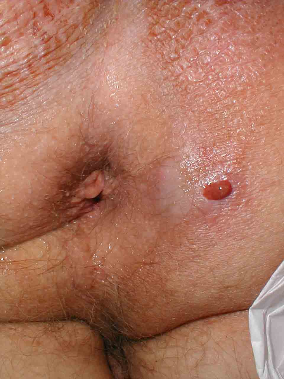
Perianal abscess
-
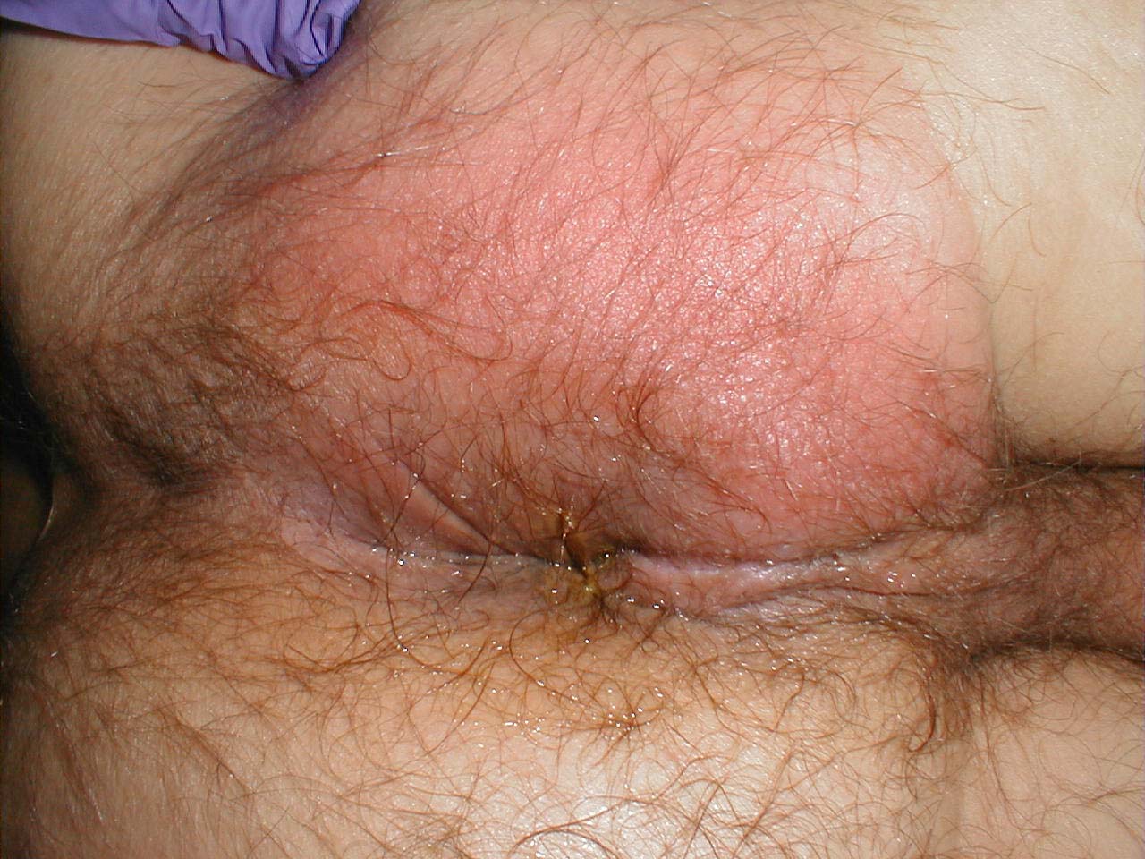
Perianal abscess


(Images courtesy of Charlie Goldberg, M.D., UCSD School of Medicine and VA Medical Center, San Diego, CA)
Laboratory Findings
Imaging
Treatment
Medical Therapy
Medical therapy is not recommended in patients with anal abscess as the antibiotics have poor penetration in to the abscess cavity and are not helpful to in treatment of the infection or wound healing.
Surgical Therapy
- Management of anal abscess should be prompt as the risk of involving the surrounding tissue resulting in perineal cellulitis and sepsis is high.[12]
- Therapy for anorectal abscess is incision and drainage and should be performed within 24 hours of presentation.
- Patients with perianal abscess and ischiorectal abscess can be treated in a outpatient setting under local anesthesia using 1% lidocaine with epinephrine (and bupivacaine for extended analgesia), is injected subcutaneously into the area affected by the abscess to provide adequate infilteration into the skin.
- Patients with loculations or large ischiorectal, intersphincteric, supralevator, or horseshoe abscesses should be admitted to the hospital and the procedure should be performed under anesthesia.
Procedure
- Under aseptic precautions and after cleaning and draping a scalpel is used to make a cruciate or elliptical incision over the area of flactulance.
- After incising the necrotic tissue is removed and loculations are broken using a hemostat.
- After the procedure the wound is packed with a gauze sponge which is removed after 24 hours.[13]
- Regular sitz bath is recommended after the surgery, it will help in local cleansing and wound healing.
Surgical Complications
- Postoperative bleeding
- Urinary retention
- Recurrence of the abscess
Prevention
Primary Prevention
Secondary Prevention
References
- ↑ 1.0 1.1 1.2 1.3 1.4 Abcarian H (2011). "Anorectal infection: abscess-fistula". Clin Colon Rectal Surg. 24 (1): 14–21. doi:10.1055/s-0031-1272819. PMC 3140329. PMID 22379401.
- ↑ Janicke DM, Pundt MR (1996). "Anorectal disorders". Emerg. Med. Clin. North Am. 14 (4): 757–88. doi:10.1016/S0733-8627(05)70278-9. PMID 8921768. Unknown parameter
|month=ignored (help) - ↑ 3.0 3.1 3.2 "Anatomy and Embryology - Springer".
- ↑ Rickard MJ (2005). "Anal abscesses and fistulas". ANZ J Surg. 75 (1–2): 64–72. doi:10.1111/j.1445-2197.2005.03280.x. PMID 15740520.
- ↑ PARKS AG (1961). "Pathogenesis and treatment of fistuila-in-ano". Br Med J. 1 (5224): 463–9. PMC 1953161. PMID 13732880.
- ↑ Ommer A, Herold A, Berg E, Fürst A, Sailer M, Schiedeck T (2012). "German S3 guideline: anal abscess". Int J Colorectal Dis. 27 (6): 831–7. doi:10.1007/s00384-012-1430-x. PMID 22362468.
- ↑ Read DR, Abcarian H (1979). "A prospective survey of 474 patients with anorectal abscess". Dis Colon Rectum. 22 (8): 566–8. PMID 527452.
- ↑ Adamo K, Sandblom G, Brännström F, Strigård K (2016). "Prevalence and recurrence rate of perianal abscess--a population-based study, Sweden 1997-2009". Int J Colorectal Dis. 31 (3): 669–73. doi:10.1007/s00384-015-2500-7. PMID 26768004.
- ↑ Adikrisna R, Udagawa M, Sugita Y, Ishii T, Okamoto H, Yabata E (2015). "[A Case of Squamous Cell Carcinoma of the Anal Canal with a Perianal Abscess]". Gan To Kagaku Ryoho. 42 (12): 2322–4. PMID 26805351.
- ↑ Ferri, Fred (2015). Ferri's clinical advisor 2015 : 5 books in 1. Philadelphia, PA: Elsevier/Mosby. ISBN 978-0323083751.
- ↑ Chang J, Mclemore E, Tejirian T (2016). "Anal Health Care Basics". Perm J. 20 (4): 74–80. doi:10.7812/TPP/15-222. PMC 5101094. PMID 27723447.
- ↑ Slauf P, Antoš F, Marx J (2014). "[Acute periproctal abscesses]". Rozhl Chir. 93 (4): 226–31. PMID 24881481.
- ↑ Smith SR, Newton K, Smith JA, Dumville JC, Iheozor-Ejiofor Z, Pearce LE; et al. (2016). "Internal dressings for healing perianal abscess cavities". Cochrane Database Syst Rev (8): CD011193. doi:10.1002/14651858.CD011193.pub2. PMID 27562822.