Rabies laboratory tests: Difference between revisions
Iqra Qamar (talk | contribs) No edit summary |
Iqra Qamar (talk | contribs) (→Biopsy) |
||
| Line 1: | Line 1: | ||
__NOTOC__{{CMG}} | __NOTOC__ | ||
{{CMG}} | |||
Please help WikiDoc by adding more content here. It's easy! Click [[Help:How_to_Edit_a_Page|here]] to learn about editing. | Please help WikiDoc by adding more content here. It's easy! Click [[Help:How_to_Edit_a_Page|here]] to learn about editing. | ||
| Line 85: | Line 87: | ||
The rabies antibody used for the dFA test is primarily directed against the [[nucleoprotein]] (antigen) of the virus. Rabies virus [[replicates]] in the [[cytoplasm]] of [[cells]], and infected cells may contain large round or oval inclusions containing collections of nucleoprotein (N) or smaller collections of [[antigen]] that appear as dust-like fluorescent particles if stained by the dFA procedure. | The rabies antibody used for the dFA test is primarily directed against the [[nucleoprotein]] (antigen) of the virus. Rabies virus [[replicates]] in the [[cytoplasm]] of [[cells]], and infected cells may contain large round or oval inclusions containing collections of nucleoprotein (N) or smaller collections of [[antigen]] that appear as dust-like fluorescent particles if stained by the dFA procedure. | ||
== References == | |||
==References== | |||
{{Reflist|2}} | {{Reflist|2}} | ||
{{WH}} | {{WH}} | ||
Revision as of 22:27, 27 September 2017
Editor-In-Chief: C. Michael Gibson, M.S., M.D. [1]
Please help WikiDoc by adding more content here. It's easy! Click here to learn about editing.
Overview
Routine lab findings are non-specific and may include
Laboratory Findings
Routine lab findings are non-specific
- Complete blood smear
- Peripheral leukocytosis (6-8% atypical monocytes)
- Arterial blood gas
- Respiratory alkalosis followed by respiratory acidosis
- Urinalysis
- Albuminuria
- Pyuria
- Lumbar puncture:
- Lymphocytic pleocytosis (mean 60 cells/uL)
- CSF protein-elevated (typically less than 100 mg/dL)
- Normal glucose concentration
Serology
- Serology is useful in assessing the serostatus in immunized humans and animals as it is difficult to document the neutralizing antibody response via immunofluorescence because mostly death occurs prior to mounting a response[1]
- The interpretation of results depends upon:[2][3]
- The specific specimen
- The history of immunization
In order to make a lab diagnosis of Rabies, certain specimens need to be taken from the body and specific tests done are as follows:
| Sample | Sensitivity | Specificity | Test | Significance/Interpretation |
|---|---|---|---|---|
| Saliva | 63.2 | 70.2 | Reverse transcriptase polymerase chain reaction (RT-PCR) | Detects viral RNA |
| Viral culture | Isolates the virus | |||
| Skin biopsy | 98 | 98.3 | Reverse transcriptase polymerase chain reaction (RT-PCR) | Detects viral RNA |
| Immunofluorescence staining | Detects viral antigen | |||
| Serum | low | low | Indirect immunofluorescence | Detects viral antigen |
| Virus neutralization assays | Detects antibody response | |||
| Cerebrospinal fluid | low | low | Indirect immunofluorescence | Detects viral antigen |
| Virus neutralization assays | Detects antibody response |
Direct Fluorescent Antibody Test (dFA)
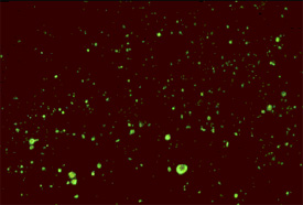
The dFA test is based on the observation that animals infected by rabies virus have rabies virus proteins (antigen) present in their tissues. Because rabies is present in nervous tissue (and not blood like many other viruses), the ideal tissue to test for rabies antigen is brain. The most important part of a dFA test is flouresecently-labeled anti-rabies antibody. When labeled antibody is incubated with rabies-suspect brain tissue, it will bind to rabies antigen. Unbound antibody can be washed away and areas where antigen is present can be visualized as fluorescent-apple-green areas using a fluorescence microscope. If rabies virus is absent there will be no staining.
The image with the bright green spots displays a positive test for rabies and the image below it, with no spots, displays a negative test for rabies.
The rabies antibody used for the dFA test is primarily directed against the nucleoprotein (antigen) of the virus. Rabies virus replicates in the cytoplasm of cells, and infected cells may contain large round or oval inclusions containing collections of nucleoprotein (N) or smaller collections of antigen that appear as dust-like fluorescent particles if stained by the dFA procedure.
References
- ↑ Manning SE, Rupprecht CE, Fishbein D, Hanlon CA, Lumlertdacha B, Guerra M, Meltzer MI, Dhankhar P, Vaidya SA, Jenkins SR, Sun B, Hull HF (2008). "Human rabies prevention--United States, 2008: recommendations of the Advisory Committee on Immunization Practices". MMWR Recomm Rep. 57 (RR-3): 1–28. PMID 18496505.
- ↑ Noah DL, Drenzek CL, Smith JS, Krebs JW, Orciari L, Shaddock J, Sanderlin D, Whitfield S, Fekadu M, Olson JG, Rupprecht CE, Childs JE (1998). "Epidemiology of human rabies in the United States, 1980 to 1996". Ann. Intern. Med. 128 (11): 922–30. PMID 9634432.
- ↑ Warrell MJ, Warrell DA (2004). "Rabies and other lyssavirus diseases". Lancet. 363 (9413): 959–69. doi:10.1016/S0140-6736(04)15792-9. PMID 15043965.

