Peritoneal dialysis: Difference between revisions

Jump to navigation Jump to search
Line 33: Line 33:


==Complications==
==Complications==
Peritoneal dialysis requires access to the [[peritoneum]]. As this access breaks normal skin barriers, and as people with renal failure generally have a slightly suppressed immune system, [[infection]] is a relatively common problem. With the development of prophylaxis therapies, infection rates have been dramatically decreased. The infections can be localized, as in an exit-site or tunnel infection, where the infection is limited to the skin or soft tissue around the catheter, or potentially more severe, if the infection reaches the peritoneum, in which case it is termed PD [[peritonitis]]; which may require [[antibiotic]]s and supportive care, or, if the peritonitis is severe, removal of the catheter and a change of [[renal replacement therapy]] modality to [[hemodialysis]]. Occasionally, severe peritonitis may be life-threatening.
* [[infection]]
Long term peritoneal dialysis can cause changes in the peritoneal membrane, making it less permeable and causing it to no longer act as a dialysis membrane as well as it used to. This loss of function can manifest as a loss of dialysis adequacy, or poorer fluid exchange (also known as '''ultrafiltration failure'''). It may also cause diabetes because of the glucose levels that are in the 'bags'
* [[peritonitis]]
 
* [[Hernias]]  
Other complications that can occur are fluid leaks into surrounding soft tissue, often the [[scrotum]] in males. [[Hernias]] are another problem that can occur due to the [[abdominal]] fluid load. These often require repair before peritoneal dialysis is recommenced.
* Accumulation of [[fibrin]] in the PD effluent  
 
* Damage to [[Peritoneal membrane.|peritoneal membrane]]
Also, a common issue that arises in PD patients is the accumulation of [[fibrin]] in the PD effluent.  This can cause draining and/or filling issues if too much collects in or around the [[catheter]] inside the [[peritoneum]].  To break up the [[fibrin]], [[Heparin]] must be injected into the bags of [[dialysate]] (generally 1mL [[Heparin]] per liter of [[dialysate]]) until the [[fibrin]] clears up.  One sign of [[peritonitis]] is the accumulation of very large amounts of [[fibrin]] in the PD effluent.
* [[Diabetes]] because of the [[glucose]] levels that are in the 'bags'
 
* [[Fluid]] leaks into surrounding [[soft tissue]], often the [[scrotum]] in males
===Differentiating CAPD from other causes of peritonitis===
 
{| style="margin: 1em 1em 1em 0; background: #f9f9f9; border: 1px #aaa solid; border-collapse: collapse;" cellspacing="0" cellpadding="4" border="2"
|+'''Differentiating SBP from other causes of peritonitis'''
! colspan="2" style="background: #4479BA; text-align: center;" | {{fontcolor|#FFF| '''Disease'''}}
! colspan="1" style="background: #4479BA; text-align: center;" | {{fontcolor|#FFF| '''Prominent clinical findings'''}}
! colspan="1" style="background: #4479BA; text-align: center;" | {{fontcolor|#FFF| '''Lab tests'''}}
! colspan="1" style="background: #4479BA; text-align: center;" | {{fontcolor|#FFF| '''Tratment'''}}
|-
| rowspan="3" |'''Primary peritonitis'''
|'''[[Primary peritonitis|Spontaneous bacterial peritonitis]]'''
|
* Absence of GI [[perforation]], most closely associated with [[cirrhosis]] and [[Liver disease|advanced liver disease]].
* Presents with abrupt onset of [[fever]], [[abdominal pain]], [[distension]], and [[rebound tenderness]].
|
* Most have clinical and biochemical manifestations of advanced [[cirrhosis]] or [[nephrosis]] like [[leukocytosis]],[[hypoalbuminemia]],
* Prolonged [[prothrombin]] time. SAAG >1.1 g/dL, increased serum [[lactic acid]] level, or a decreased [[Ascites|ascitic fluid]] pH (< 7.31) supports the diagnosis. [[Gram staining]] reveals bacteria in only 25% of cases.
* Diagnosed by analysis of the [[Ascitic|ascitic fluid]] which reveals [[WBC]] > 500/ML, and [[PMN]] >250cells/ml.
* [[Culture medium|Culture]] of ascitic fluid inoculated immediately into [[blood culture]] media at the bedside usually reveals a single [[Enteric Bacilli|enteric organism]], most commonly ''[[Escherichia coli]]'', ''[[Klebsiella]]'', or [[streptococci]].
|
* Once diagnosed,it is treated with [[Ceftriaxone]].
|-
|'''[[Tuberculous peritonitis]]'''
|
* Seen in 0.5% of new cases of [[tuberculosis]] particularly in young women in endemic areas as a primary infection.
* Presents with [[abdominal pain]] and [[distension]], [[fever]], [[night sweats]], [[weight loss]], and altered bowel habits.
|
* [[Ascites]] is present in about half of cases. [[Abdominal mass]] may be felt in a third of cases. The [[peritoneal fluid]] is characterized by a [[protein]] concentration > 3 g/dL with < 1.1 g/dL SAAG and [[Lymphocyte|lymphocyte predominance]] of [[WBC]].
* Definitive diagnosis in 80% of cases is by culture. Most patients presenting acutely are diagnosed only by [[laparotomy]].
|
* Combination [[Antituberculosis|antituberculosis chemotherapy]] is preferred in chronic cases.
|-
|'''[[Continuous ambulatory peritoneal dialysis|Continuous Ambulatory Peritoneal Dialysis]]''' [[Continuous ambulatory peritoneal dialysis|('''CAPD peritonitis)''']]
|
* [[Peritonitis]] is one of the major complications of [[peritoneal dialysis]] & 72.6% occurred within the first six months of [[peritoneal dialysis]].
* Historically, [[coagulase-negative staphylococci]] were the most common cause of peritonitis in [[Continuous ambulatory peritoneal dialysis|CAPD]], presumably due to touch contamination or infection via the pericatheter route.
* Treatment for [[peritoneal dialysis]]-associated peritonitis consists of [[Antimicrobial drug|antimicrobial therapy]], in some cases catheter removal is also warranted.
* Additional therapies for [[Peritonitis|relapsing or recurrent peritonitis]] may include [[Fibrinolytic agent|fibrinolytic agents]] and [[peritoneal lavage]]. Most episodes of peritoneal dialysis-associated peritonitis resolve with outpatient [[Antibiotic|antibiotic treatment]].
|
* Majority of [[peritonitis]] cases are caused by [[bacteria]] (50%-due to [[Gram-positive bacteria|gram positive]] organisms, 15% to [[gram negative]] organisms,20% were culture negative.2% of cases are caused by [[fungi]], mostly [[Candida]] species. Polymicrobial infection in 4%.Exit-site infection was present in 13% and a [[peritoneal fluid]] leak in 3 % and [[M.tuberculosis]] 0.1%.
|
* [[Antibiotic|Initial empiric antibiotic coverage]] for peritoneal dialysis-associated peritonitis consists of coverage for [[gram-positive]] organisms (by [[vancomycin]] or a [[Cephalosporins|first-generation cephalosporin]]) and [[gram-negative]] organisms (by a [[cephalosporin|third-generation cephalosporin]] or an [[aminoglycoside]]). Subsequently, the regimen should be adjusted based on [[Culture medium|culture]] and [[sensitivity]] data. Cure rates are approximately 75%.
|-
| rowspan="2" |'''[[Secondary peritonitis]]'''
|'''Acute [[bacterial]] [[secondary peritonitis]]'''
|
* Occurs after perforating, penetrating, inflammatory, infectious, or [[ischemic]] injuries of the GI or GU tracts. Most often follows disruption of a hollow viscus?chemical peritonitis?bacterial peritonitis(polymicrobial, includes [[aerobic]] [[gram negative]] {[[E coli]], [[Klebsiella]], [[Enterobacter]], [[Proteus mirabilis]]} and gram positive { [[Enterococcus]], [[Streptococcus]]} and [[anaerobes]] {[[Bacteroides]], [[clostridia]]}).
* Presents with [[abdominal pain]], [[tenderness]], [[guarding]] or rigidity, [[distension]], free peritoneal air, and diminished [[bowel sounds]]. Signs that reflect irritation of the parietal peritoneum resulting [[ileus]]. Systemic findings include [[fever]], [[chills]] or [[rigors]], [[tachycardia]], [[sweating]], [[tachypnea]], [[restlessness]], [[dehydration]], [[oliguria]], [[disorientation]], and, ultimately, refractory [[shock]].
|
|
* [[Peritoneal lavage]], [[Laparoscopy]] are the treatment of choice.
|-
|'''[[Biliary]] [[Secondary peritonitis|peritonitis]]'''
|
* Most often seen in cases of rupture of pathological [[gallbladder]] or [[bile duct]] or [[Cholangitis|cholangitic abscess]] or secondary to obstruction of  the [[biliary tract]].
* Seen in alcoholic patients with [[ascites]].
|
|
|-
| colspan="2" |'''[[Peritonitis|Tertiary peritonitis]]'''
|
* Persistence or recurrence of [[Infection|intraabdominal infection]] following apparently adequate therapy of [[Peritonitis|primary or secondary peritonitis]].
* Associated with [[Mortality|high mortality]] due to multi organ dysfunction. It presents in a similar way as other [[peritonitis]] but is recognized as an adverse outcome with poor prognosis.
|
* [[Enterococcus]], [[Candida]], [[Staphylococcus epidermidis]], and [[Enterobacter]] being the most common organisms.
|
* Characterized by lack of response to appropriate surgical and [[antibiotic therapy]] due to disturbance in the hosts [[immune response]].
|-
| colspan="2" |'''[[Familial mediterranean fever|Familial Mediterranean fever (periodic peritonitis, familial paroxysmal polyserositis)]]'''
|
* Rare [[Genetic disorder|genetic condition]] which affects individuals of Mediterranean genetic background.
* Etiology is unclear.
* Presents with recurrent bouts of [[abdominal pain]] and [[tenderness]] along with [[pleuritic]] or [[joint pain]]. [[Fever]] and [[leukocytosis]] are common.
|
|
* [[Colchicine]] prevents but does not treat acute attacks.
|-
| colspan="2" |'''[[Granulomatous peritonitis]]'''
|
* A rare condition caused by disposable surgical fabrics or food particles from a [[perforated ulcer]], eliciting a vigorous [[granulomatous]] ([[Hypersensitivity|delayed hypersensitivity]]) response in some patients 2-6 weeks after [[laparotomy]].
* Presents with [[abdominal pain]], [[fever]], [[nausea and vomiting]], [[ileus]], and systemic complaints, mild and diffuse [[abdominal tenderness]].
|
* Diagnosed by the demonstration of diagnostic Maltese cross pattern of starch particles.
|
* The disease is self-limiting.
* Treated with [[corticosteroids]] or [[Anti inflammatory medications|anti-inflammatory agents]].
|-
| colspan="2" |'''[[Sclerosing encapsulating peritonitis]]'''
|
* Seen in conditions associated with long term [[peritoneal dialysis]], shunts like [[Ventriculoperitoneal shunt|VP shunts]], history of [[Abdominal surgery|abdominal surgeries]], [[liver transplantation]].
* Symptoms include [[nausea]], [[abdominal pain]], [[diarrhea]], [[anorexia]], bloody [[ascites]].
|
|
|-
| colspan="2" |'''[[Abscess|Intraperitoneal abscesses]]'''
|
* Most common etiologies being [[Perforation|Gastrointestinal perforations]], postoperative complications, and penetrating injuries.
* Signs and symptoms depend on the location of the [[abscess]] within the [[peritoneal cavity]] and the extent of involvement of the surrounding structures.
* Diagnosis is suspected in any patient with a predisposing condition. In a third of cases it occurs as a sequela of [[Peritonitis|generalized peritonitis]].
* The pathogenic organisms are similar to those responsible for [[peritonitis]], but [[anaerobic]] organisms occupy an important role.
* The [[mortality rate]] of serious [[Abscesses|intra-abdominal abscesses]] is about 30%.
|
* Diagnosed best by [[CT-scans|CT]] scan of the abdomen.
|
* Treatment consists of prompt and complete [[CT]] or [[Ultrasound|US]] guided drainage of the [[abscess]], control of the primary cause, and adjunctive use of effective [[Antibiotics|antibiotics.]] Open drainage is reserved for [[abscesses]] for which percutaneous drainage is inappropriate or unsuccessful.
|-
| colspan="2" |'''[[Peritoneal mesothelioma]]'''
|
* Arises from the [[mesothelium]] lining the [[peritoneal cavity]].
* Its incidence is approximately 300-500 new cases being diagnosed in the United States each year.  As with [[pleural mesothelioma]], there is an association with an [[Asbestos|asbestos exposure]].
* Most commonly affects men at the age of 50-69 years. Patients most often present with [[abdominal pain]] and later increased abdominal girth and [[ascites]] along with [[anorexia]], [[weight loss]] and [[abdominal pain]].
* Mean time from diagnosis to death is less than 1 year without treatment. 
|
* [[Computed tomography|CT]] with [[Contrast|intravenous contrast]] typically demonstrates the thickening of the [[peritoneum]]. [[Laparoscopy]] with tissue biopsy or CT guided tissue biopsy with [[immunohistochemical staining]] for [[calretinin]], [[cytokeratin|cytokeratin 5/6]], [[mesothelin]], and [[WT1|Wilms tumor 1 antigen]] remain the [[Gold standard (test)|gold standard]] for diagnosis.
|
* At [[laparotomy]] the goal is cytoreduction with [[excision]]. Debulking surgery and intraperitoneal [[chemotherapy]] improves survival in some cases.
|-
| colspan="2" |'''[[peritoneal carcinomatosis]]'''
|
* Associated with a history of [[ovarian]] or [[Malignancy|GI tract malignancy]].
* Symptoms include [[ascites]], [[abdominal pain]], [[nausea]], [[vomiting]].
|
|
|}


==Step-by-step description of Peritoneal dialysis (a CAPD exchange)==

Revision as of 19:01, 27 June 2018

Peritoneal dialysis

WikiDoc Resources for Peritoneal dialysis

Articles

Most recent articles on Peritoneal dialysis

Most cited articles on Peritoneal dialysis

Review articles on Peritoneal dialysis

Articles on Peritoneal dialysis in N Eng J Med, Lancet, BMJ

Media

Powerpoint slides on Peritoneal dialysis

Images of Peritoneal dialysis

Photos of Peritoneal dialysis

Podcasts & MP3s on Peritoneal dialysis

Videos on Peritoneal dialysis

Evidence Based Medicine

Cochrane Collaboration on Peritoneal dialysis

Bandolier on Peritoneal dialysis

TRIP on Peritoneal dialysis

Clinical Trials

Ongoing Trials on Peritoneal dialysis at Clinical Trials.gov

Trial results on Peritoneal dialysis

Clinical Trials on Peritoneal dialysis at Google

Guidelines / Policies / Govt

US National Guidelines Clearinghouse on Peritoneal dialysis

NICE Guidance on Peritoneal dialysis

NHS PRODIGY Guidance

FDA on Peritoneal dialysis

CDC on Peritoneal dialysis

Books

Books on Peritoneal dialysis

News

Peritoneal dialysis in the news

Be alerted to news on Peritoneal dialysis

News trends on Peritoneal dialysis

Commentary

Blogs on Peritoneal dialysis

Definitions

Definitions of Peritoneal dialysis

Patient Resources / Community

Patient resources on Peritoneal dialysis

Discussion groups on Peritoneal dialysis

Patient Handouts on Peritoneal dialysis

Directions to Hospitals Treating Peritoneal dialysis

Risk calculators and risk factors for Peritoneal dialysis

Healthcare Provider Resources

Symptoms of Peritoneal dialysis

Causes & Risk Factors for Peritoneal dialysis

Diagnostic studies for Peritoneal dialysis

Treatment of Peritoneal dialysis

Continuing Medical Education (CME)

CME Programs on Peritoneal dialysis

International

Peritoneal dialysis en Espanol

Peritoneal dialysis en Francais

Business

Peritoneal dialysis in the Marketplace

Patents on Peritoneal dialysis

Experimental / Informatics

List of terms related to Peritoneal dialysis

Editor-In-Chief: C. Michael Gibson, M.S., M.D. [1]

Overview

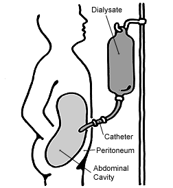
Peritoneal dialysis is a method for removing waste such as urea and potassium from the blood, as well as excess fluid, when the kidneys are incapable of this (i.e. in renal failure). It is a form of renal dialysis, and is thus a renal replacement therapy.

Peritoneal dialysis works on the principle that the peritoneal membrane that surrounds the intestine, can act as a natural semipermeable membrane (see dialysis), and that if a specially formulated dialysis fluid is instilled around the membrane then dialysis can occur, by diffusion. Excess fluid can also be removed by osmosis, by altering the concentration of glucose in the fluid.

Dialysis fluid is instilled via a peritoneal dialysis catheter, (the most common type is called a Tenckhoff Catheter) which is placed in the patient's abdomen, running from the peritoneum out to the surface, near the navel. Peritoneal dialysis catheters may also be tunneled under the skin and exit alternate locations such as near the rib margin or sternum (called a presternal catheter), or even up near the clavicle. This is done as a short surgery. The exit site is chosen based on surgeon's or patient's preference and can be influenced by anatomy or hygiene issues.

Peritoneal dialysis is typically done in the patient's home and workplace, but can be done almost anywhere; a clean area to work, a way to elevate the bag of dialysis fluid and a method of warming the fluid are all that is needed. The main consideration is the potential for infection. Peritonitis is the most common serious complication, but with good technique can usually be avoided. Infections of the catheter's exit site or "tunnel" (path from the peritoneum to the exit site) are less serious. Because of this, patients are advised to take a number of precautions against infection.

Types of peritoneal dialysis

There are three types of peritoneal dialysis.

  • Continuous ambulatory peritoneal dialysis (CAPD), the most common type, needs no machine and can be done at home. Exchanges of fluid are done throughout the day, usually four exchanges a day.
  • Continuous cyclic peritoneal dialysis (CCPD) uses a machine and is usually performed at night when the person is sleeping.
  • Intermittent peritoneal dialysis (IPD) uses the same type of machine as CCPD - if done overnight is called Nocturnal intermittent peritoneal dialysis (NIPD).

Advantages and disadvantages of Peritoneal dialysis

Advantages

  • Can be done at home.
  • Relatively easy to learn.
  • Easy to travel with, bags of solution are easy to take on holiday.
  • Fluid balance is usually easier than on hemodialysis
  • Theoretically better to start dialysis on, as native urine output is maintained for longer than on hemodialysis.
  • Excluding kidney transplant, PD is method of the first choice in treating chronic kidney failure.

Disadvantages

  • Requires a degree of motivation and attention to cleanliness while performing exchanges.
  • Possible complications (see below)

Complications

Step-by-step description of Peritoneal dialysis (a CAPD exchange)

  1. The supplies and materials needed for an exchange are...VEXORG THE DEVOURER OF CHICKENS and STUFF. Notable amongst these is a bag of dialysis fluid (also called dialysis solution), a solution comprised of a known amount of a glucose dissolved in water. The strength of this solution determines the osmotic gradient, and therefore the amount of water that diffuses out of the bloodstream. Common strengths of glucose are 0.5%, 1.5%, 2.5% and 4.25%. 1.5% is approximately fluid-neutral; it neither adds nor removes fluid and is used for patients who are primarily concerned with waste removal rather than fluid regulation. Higher concentrations lead to greater water removal. A higher dextrose concentration moves fluid and more wastes into the abdominal cavity, increasing both early and long-dwell exchange efficiency. Eventually, however, the body absorbs dextrose from the solution. As the concentration of dextrose in the body comes closer to that in the solution, dialysis becomes less effective, and fluid is slowly absorbed from the abdominal cavity. Electrolytes are also present in the fluid to maintain proper body levels. Patients weigh themselves, and measure temperature and blood pressure daily to determine whether the body is retaining fluid and, thus, what strength of fluid to use. Dialysis fluid typically comes premixed in a disposable bag-and-tube apparatus; no additional equipment is needed. The apparatus consists of two bags, one empty and one with the fluid, connected via flexible tubing to a Y-shaped fitting. The bag is heated to body temperature, to avoid causing cramping. Dry heat is used; common methods include microwaves (microwave heating is NOT recommended), heating pads and solar radiation (often using the dashboard of a car, for instance while travelling).
  2. The patient, who performs the entire procedure themselves, dons a disposable surgical mask, scrubs their hands using antibacterial soap, and tucks a clean towel into the waistband of their pants to protect their clothing. The bag of dialysis fluid is removed from the protective packaging, and is hung from an IV stand or other elevated location, such as a coat hook. The tubing attached to the bag of fluid is uncoiled, and the second (empty) bag is placed on the floor. The Y-shaped connector is attached to the catheter tip; a protective cap must be removed from both of these before the connection is made, and the two portions of the connector are not permitted to touch anything, to avoid possible contamination.
  3. Once connected to the system, the patient clamps the tubing connected to the full bag of dialysis fluid and then releases the twist valve located in the tip of their catheter; this permits fluid to flow into or out of the peritoneal cavity. Because the full bag of fluid is clamped off but the empty bag is not, the effluent (used dialysis fluid) from within the peritoneum can drain out of the catheter and into the lower, waste bag. Emptying the abdomen of fluid takes approximately fifteen minutes, and the patient is free to perform tasks such as reading, watching television and browsing the internet.
  4. When the abdomen has drained, the lower drain-bag is clamped off. The twist valve in the catheter is also closed. The clamp is then removed from the upper tubing, permitting dialysis fluid to drain out into the abdomen. The clamp to the drain bag is briefly opened and some fluid is drained directly from the upper bag into the lower bag. This clears the line of air and other impurities. The drain line is then clamped off and the twist valve on the catheter end is opened. This permits fluid to enter the peritoneum. Filling the abdomen with fresh fluid takes about fifteen minutes, and the patient enjoys the same freedoms as while draining.
  5. Once the entire bag of fluid (an amount varying primarily based on body size, ranging from 1500 to 3000 mL) has been introduced to the abdomen, the patient then cleans their hands again (typically using an antiseptic alcohol-based cleanser) and puts the surgical mask on. The Y-connector is detached from the catheter tip and a protective cap is placed on the end of the catheter.
  6. The effluent is inspected after a dialysis exchange is complete; a cloudy effluent indicates probable peritoneal infection. The effluent is drained into a toilet, and the various dialysis supplies are discarded with normal garbage.

See also

Template:WH Template:WS