Osteosarcoma pathophysiology
Editor-In-Chief: C. Michael Gibson, M.S., M.D. [1]
|
Osteosarcoma Microchapters |
|
Diagnosis |
|---|
|
Treatment |
|
Case Studies |
|
Osteosarcoma pathophysiology On the Web |
|
American Roentgen Ray Society Images of Osteosarcoma pathophysiology |
|
Risk calculators and risk factors for Osteosarcoma pathophysiology |
Overview
Pathophysiology
The tumor may be localized at the end of the long bones. Most often it affects the upper end of tibia or humerus, or lower end of femur. The tumor is solid, hard, irregular ("fir-tree" or "sun-burst" appearance on X-ray examination) due to the tumor spicules of calcified bone radiating in right angles. These right angles form what is known as Codman's triangle. Surrounding tissues are infiltrated.
Microscopically: The characteric feature of osteosarcoma is presence of osteoid (bone formation) within the tumour. Tumor cells are very pleomorphic (anaplastic), some are giant, numerous atypical mitoses. These cells produce osteoid describing irregular trabeculae (amorphous, eosinophilic/pink) with or without central calcification (hematoxylinophilic/blue, granular) - tumor bone. Tumor cells are included in the osteoid matrix. Depending on the features of the tumour cells present (whether they resemble bone cells, cartilage cells or fibroblast cells), the tumour can be subclassified. Presence of immature blood vessels (sarcomatous vessels lacking endothelial cells) favors the bloodstream metastasizing.
Genetics
Hereditary syndromes of osteosarcoma have been identified[1]:
- Rothmund-Thomson Syndrome
- RECQL4 gene mutations
- RB1 gene mutations (also implicated in retinoblastoma)
- Li-Fraumeni syndrome
These syndromes are extremely rare within the Osteosarcoma diagnosis, and probably represent less than 0.5% of those diagnosed.
-
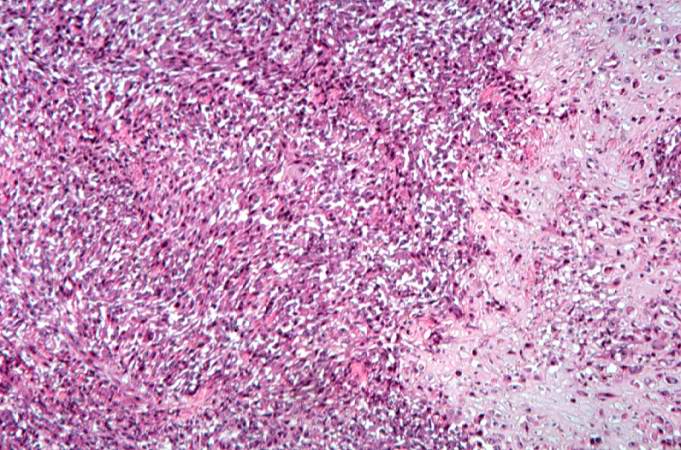
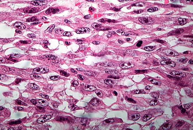
This high-power photomicrograph demonstrates the cellular growth pattern. Note that the cells are fusiform and they grow in sheets.
-
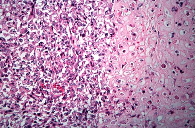
This high-power photomicrograph demonstrates the growth pattern and the cell morphology.
-
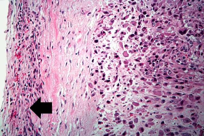
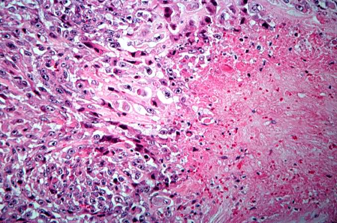
This is a high-power photomicrograph of the tumor cell morphology and the periosteum (arrow).
-
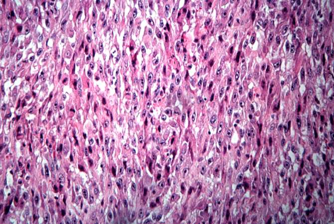
This high-power photomicrograph of the tumor demonstrates the fusiform morphology of the cells. Note the marked variability in size and staining intensity of the nuclei.
-
This is a high-power photomicrograph of the tumor demonstrating the anaplastic cell morphology.
-
This is a high-power photomicrograph of the tumor demonstrating the anaplastic cell morphology.
-
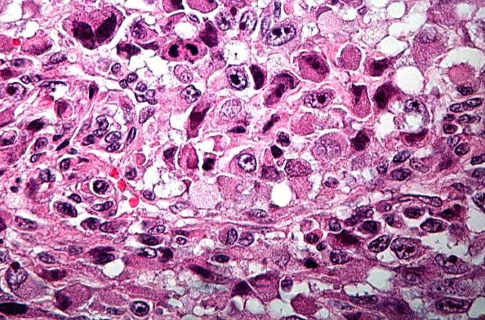
This is a high-power photomicrograph of the tumor demonstrating the anaplastic cell morphology and multiple mitotic figures (arrows).


References
- ↑ Wang LL. Biology of osteogenic sarcoma. Cancer J 11:294-305, 2005.