OctreoScan: Difference between revisions
Jump to navigation
Jump to search
(New page: right|300px ==Indications for imaging== *Tumor evaluation (i.e. Pheochromocytoma , Paraganglioma , Islet cell tumor , Carcinoid , [[Me...) |
No edit summary |
||
| Line 3: | Line 3: | ||
==Indications for imaging== | ==Indications for imaging== | ||
*Tumor evaluation | * Tumor evaluation | ||
:* [[Pheochromocytoma]] | |||
:* [[Paraganglioma]] | |||
:* [[Islet cell tumor]] | |||
:* [[Carcinoid]] | |||
:* [[Medullary carcinoma of thyroid]] | |||
:* [[Small cell lung cancer]] | |||
:* [[Neuroblastoma]] | |||
:* [[Pituitary adenoma]] | |||
:* [[Lymphoma]] | |||
:* [[Breast cancer]] | |||
:* [[Astrocytoma]] | |||
:* [[Meningioma]] | |||
:* [[Thymoma]] | |||
==Patient preparation== | ==Patient preparation== | ||
*Bowel preparation with laxative and enema | * Bowel preparation with laxative and enema | ||
*Discontinue octeotide therapy for 1 week prior the exam | * Discontinue octeotide therapy for 1 week prior the exam | ||
==Radiopharmaceutical== | ==Radiopharmaceutical== | ||
Revision as of 16:53, 26 February 2009
Indications for imaging
- Tumor evaluation
Patient preparation
- Bowel preparation with laxative and enema
- Discontinue octeotide therapy for 1 week prior the exam
Radiopharmaceutical
- In-111 OctreoScan
Dose and route of administration
- 6 mCi IV
- 4 hours (abdomen, pelvis)
- 24 hours (whole body)
Equipment
- Camera: Large field of view SPECT gamma camera
- Collimator: Medium-energy
- Window: 20% window around 173 and 245 KeV
Procedure
- 4 hours: Planar images of the abdomen and pelvis
- 24 hours: Planar images of the whole body. SPECT of regions of clinical concern.
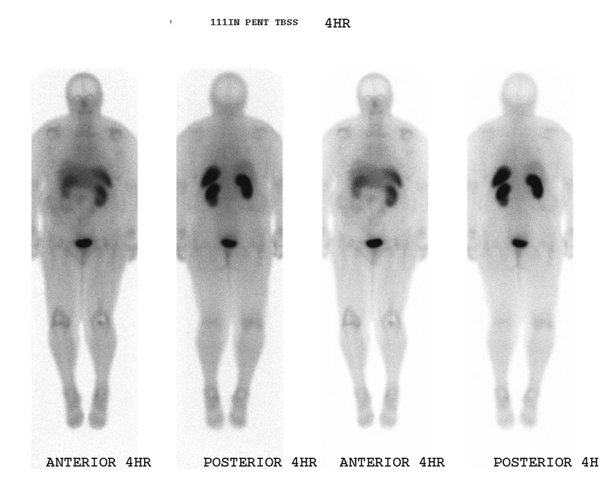
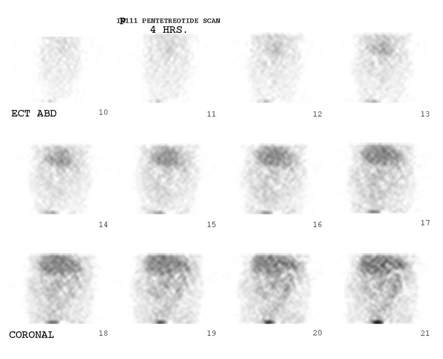
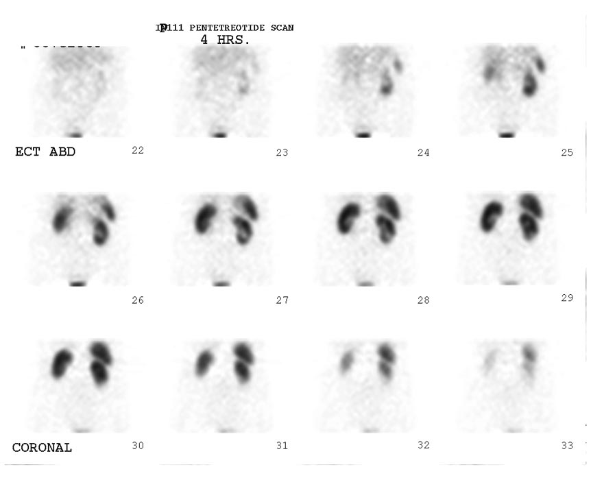
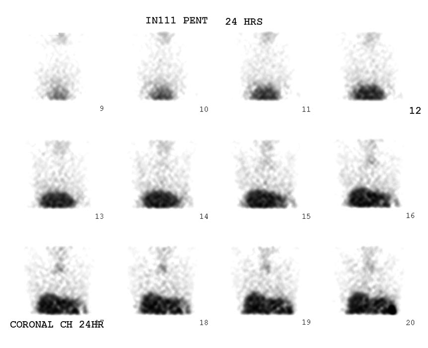
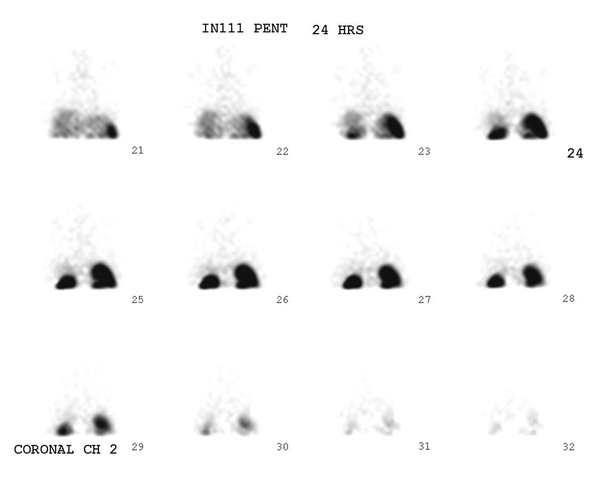
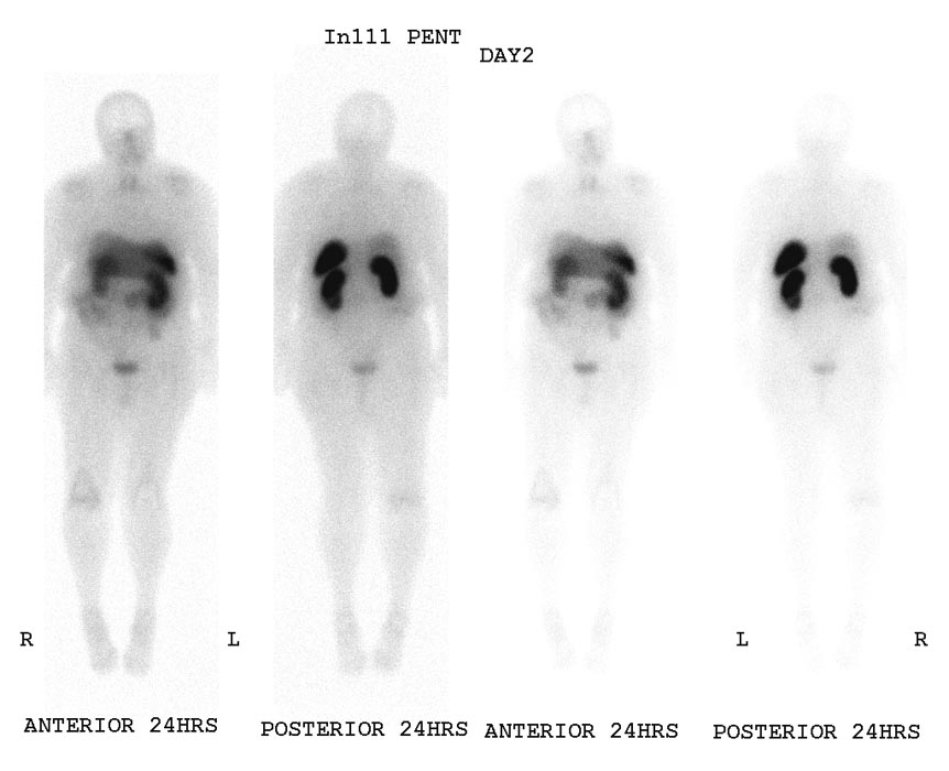
Images
Normal OctreoScan
References
- Ziessman, Harvey A, O'Malley, Janis P, and Thrall, James M. Nuclear Medicine: The Requisites. 3rd ed. Philadelphia, Pennsylvania: Mosby, 2006. ISBN 0323029469
