Nasopharyngeal angiofibroma
| Nasopharyngeal angiofibroma | |
 | |
|---|---|
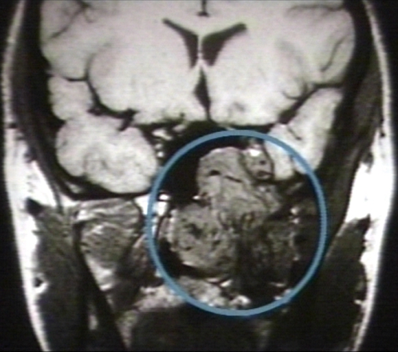
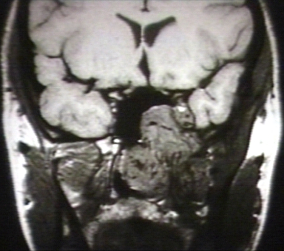
| MRI (T1): Nasopharyngeal angiofibroma. Image courtesy of Professor Peter Anderson DVM PhD and published with permission © PEIR, University of Alabama at Birmingham, Department of Pathology | |
| DiseasesDB | 32229 |
| MedlinePlus | 001572 |
| eMedicine | ent/470 |
| MeSH | D018322 |
Editor-In-Chief: C. Michael Gibson, M.S., M.D. [1]
Contributors: Cafer Zorkun M.D., PhD.
Overview
Nasopharyngeal angiofibroma (also called juvenile nasopharyngeal angiofibroma) is a histologically benign but locally aggressive vascular tumor that grows in the back of the nasal cavity. It almost exclusively affects adolescent males. Patients with nasopharyngeal angiofibroma usually present with one-sided nasal obstruction and recurrent bleeding.
Differential Diagnosis
- Antro-choanal polyp (antral-choanal polyp)
- Rhinosporidiosis
- Malignancy
- Chordoma
- Nasopharanageal cyst
- Pyogenic granuloma
Diagnosis
Symptoms
- Frequent epistaxis or blood-tinged nasal discharge
- Nasal obstruction and rhinorrhea
- Conductive hearing loss from eustachian-tube obstruction
- Diplopia, which occurs secondary to erosion into the cranial cavity and pressure on the optic chiasma
- Rarely anosmia, recurrent otitis media, and eye pain
CT and MRI
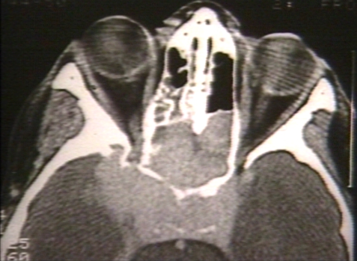
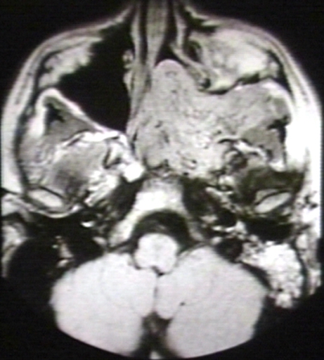
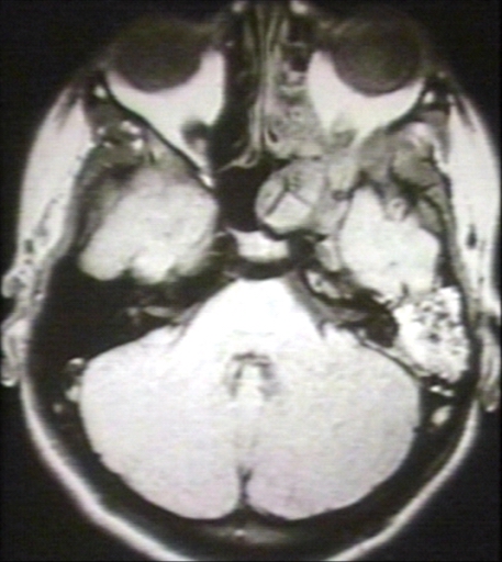
If nasopharyngeal angiofibroma is suspected based on physical exam (a smooth submucosal mass in the posterior nasal cavity), imaging studies such as CT or MRI should be performed.

Biopsy
Biopsy can lead to extensive bleeding since the tumor is composed of blood vessels without a muscular coat.
Treatment
Nasopharyngeal angiofibroma is treated by surgery. Medical treatment is usually given before surgery to reduce the blood loss - includes the usage of Diethylstilbestrol 2-3 weeks before surgery to make the surgery relatively less vascular.
Pre-surgery angiography may allow for embolization, reducing intraoperative blood loss. Hypotensive anaesthesia is the usual modality followed to reduce blood loss. Patients with tumors that have extended into the cranial cavity or whose tumors can't be safely reached by surgery may receive radiation therapy. Also radiation therapy helps in case where recurrence is the main problem.
Endoscopy has recently been used in patients with tumors of limited extension that have been pre-operatively embolized. Then, lateral rhinotomy is the usual preferred technique these days, an incision along the lateral border of nose is required to explore the maxillary antrum and remove the pathology. Other useful method is degloving technique which requires adequate operator skills.
References
- Richard R. Orlandi. Nose and sinuses from Oxford Textbook of Surgery - 2nd Ed. Peter J. Morris and William C. Wood editors, Oxford University Press, Oxford, UK, 2000.
- Douglas R, Wormald P (2006). "Endoscopic surgery for juvenile nasopharyngeal angiofibroma: where are the limits?". Curr Opin Otolaryngol Head Neck Surg. 14 (1): 1–5. PMID 16467630.