Myxoma chest x ray: Difference between revisions
No edit summary |
|||
| (20 intermediate revisions by 5 users not shown) | |||
| Line 1: | Line 1: | ||
__NOTOC__ | __NOTOC__ | ||
{{Myxoma}} | {{Myxoma}} | ||
{{CMG}}; '''Associate Editor-In-Chief:''' {{CZ}} {{AAM}} | {{CMG}}; '''Associate Editor-In-Chief:''' {{MV}} {{CZ}} {{AAM}} | ||
==Overview== | ==Overview== | ||
There are no specific chest [[x-ray]] findings associated with cardiac myxoma, the results can be reported as normal. | |||
==Key Chest X-Ray Findings in Cardiac Myxoma == | |||
*There are no specific chest [[x-ray]] findings associated with cardiac myxoma, the results can be reported as normal.<ref> Cardiac Myxoma. Radiopedia.http://radiopaedia.org/articles/cardiac-myxoma Accessed on November 24, 2015 </ref> | |||
There are no specific chest x-ray findings associated with cardiac myxoma, the results can be reported as normal. <ref>Cardiac Myxoma. Radiopedia.http://radiopaedia.org/articles/cardiac-myxoma Accessed on November 24, 2015 </ref> | *Related imaging findings include [[cardiomegaly]], [[left atrial enlargement]], vascular redistribution, prominent [[pulmonary trunk]], and intracardiac [[tumoral]] [[calcification]] (rare).<ref name="pmid">{{cite journal |vauthors=Thyagarajan B, Kumar MP, Patel S, Agrawal A |title=Extracardiac manifestations of atrial myxomas |journal=J Saudi Heart Assoc |volume=29 |issue=1 |pages=37–43 |date=January 2017 |pmid= |pmc=5247297 |doi=10.1016/j.jsha.2016.07.003 |url=}}</ref> | ||
==Gallery== | |||
<div align=" | <div align="center"> | ||
<gallery heights="225" widths="225"> | <gallery heights="225" widths="225"> | ||
Image:Myxoma chest x-ray 1.jpg|Lateral chest radiograph from a 16-year-old girl with syncope and bacterial endocarditis. The radiograph demonstrates two areas of dense calcification (arrowheads) overlying the posterior aspect of heart. The posterior-anterior (PA) view confirmed location in the heart (not shown). At surgery a calcified myxoma of the right atrium was removed. <small>[http://www.peir.net Image courtesy of Professor Peter Anderson DVM PhD and published with permission © PEIR, University of Alabama at Birmingham, Department of Pathology]</small> | Image:Myxoma chest x-ray 1.jpg|Lateral chest radiograph from a 16-year-old girl with syncope and bacterial endocarditis. The radiograph demonstrates two areas of dense calcification (arrowheads) overlying the posterior aspect of heart. The posterior-anterior (PA) view confirmed location in the heart (not shown). At surgery a calcified myxoma of the right atrium was removed. <small>[http://www.peir.net Image courtesy of Professor Peter Anderson DVM PhD and published with permission © PEIR, University of Alabama at Birmingham, Department of Pathology]</small> | ||
| Line 17: | Line 18: | ||
</div> | </div> | ||
{| style="border: 0px; font-size: 90%; margin: 3px;" align=center | |||
|+ | |||
! style="background: #4479BA; width: 100px;" | {{fontcolor|#FFF|Imaging Technique}} | |||
! style="background: #4479BA; width: 300px;" | {{fontcolor|#FFF|Features}} | |||
! style="background: #4479BA; width: 200px;" | {{fontcolor|#FFF|Description}} | |||
! style="background: #4479BA; width: 200px;" | {{fontcolor|#FFF|Advantages}} | |||
! style="background: #4479BA; width: 300px;" | {{fontcolor|#FFF|Limitations}} | |||
|- | |||
| style="padding: 5px 5px; background: #DCDCDC;" align=center|'''Two- or three-dimensional echocardiography''' | |||
| style="padding: 5px 5px; background: #F5F5F5;" align=center| | |||
*Echocardiography is usually the initial modality used for identification and evaluation of cardiac myxomas. | |||
| style="padding: 5px 5px; background: #F5F5F5;" align=center| | |||
*Hyperechogenic lesions with a well-defined stalk. | |||
*Protrusion into the ventricles is a common finding. | |||
| style="padding: 5px 5px; background: #F5F5F5;" align=center| | |||
*Real-time imaging | |||
*Tumor mobility and distensibility. | |||
| style="padding: 5px 5px; background: #F5F5F5;" align=center| | |||
*Limited views of the [[mediastinum]] and cannot be used to evaluate extracardiac manifestations of disease.<ref name="pmid1943240">{{cite journal |vauthors=Reeder GS, Khandheria BK, Seward JB, Tajik AJ |title=Transesophageal echocardiography and cardiac masses |journal=Mayo Clin. Proc. |volume=66 |issue=11 |pages=1101–9 |year=1991 |pmid=1943240 |doi= |url=}}</ref> | |||
*[[Transesophageal echocardiography (TEE)|TEE]] is an invasive imaging technique. | |||
*[[TTE]] is limited by the imaging window, which can vary with the patient and operator experience. | |||
|- | |||
| style="padding: 5px 5px; background: #DCDCDC;" align=center|'''MRI''' | |||
| style="padding: 5px 5px; background: #F5F5F5;" align=center| | |||
*Evaluation of cardiac masses and is of greatest value when echocardiographic findings are suboptimal or when the lesion has an atypical location or appearance. | |||
| style="padding: 5px 5px; background: #F5F5F5;" align=center| | |||
*Cardiac myxomas appear spherical or ovoid with lobular contours, irregular in shape. | |||
*'''T1''' : Low to intermediate signal, but areas of hemorrhage may be high. | |||
*'''T1 C+ (Gd)''': shows enhancement (important discriminator from a thrombus) demonstrates uniform heterogeneous enhancement. | |||
| style="padding: 5px 5px; background: #F5F5F5;" align=center| | |||
* MRI allows imaging in multiple planes. | |||
* Provides some functional information such as, flow direction and flow velocity in large vessels. | |||
| style="padding: 5px 5px; background: #F5F5F5;" align=center| | |||
*Cannot show [[calcification]]. | |||
*High susceptibility to motion [[artifact]]. | |||
*Dependent on regular electrocardiographic rhythms and cardiac gating. | |||
|- | |||
| style="padding: 5px 5px; background: #DCDCDC;" align=center|'''CT''' | |||
| style="padding: 5px 5px; background: #F5F5F5;" align=center| | |||
*CT can be used to accurately image the heart and surrounding mediastinum. | |||
| style="padding: 5px 5px; background: #F5F5F5;" align=center| | |||
*Intracardiac heterogeneously low attenuating mass. | |||
*The attenuation is usually lower than that of myocardium. | |||
*[[Calcification]] is common | |||
| style="padding: 5px 5px; background: #F5F5F5;" align=center| | |||
*CT provides better soft-tissue contrast. | |||
| style="padding: 5px 5px; background: #F5F5F5;" align=center| | |||
*There is no real-time true imaging with CT and imaging planes are limited to those allowed by angulation of the gantry. | |||
*There is no evaluation of small moving structures, such as the cardiac valves. | |||
|- | |||
| style="padding: 5px 5px; background: #DCDCDC;" align=center|'''Angiography''' | |||
| style="padding: 5px 5px; background: #F5F5F5;" align=center| | |||
*Coronary angiography may be helpful to detect vascular supply of the tumor by the coronary arteries. | |||
| style="padding: 5px 5px; background: #F5F5F5;" align=center| | |||
*The angiographic findings of cardiac myxoma demonstrate feeding vessels, contrast medium poolings, and clusters of [[tortuous]] vessels that correspond to tumor vasculature | |||
| style="padding: 5px 5px; background: #F5F5F5;" align=center| | |||
*Angiography can detect the concomitant coronary disease and the unique vascular appearances of cardiac myxoma. | |||
*Helpful for surgical evaluation. | |||
| style="padding: 5px 5px; background: #F5F5F5;" align=center| | |||
*Invasive imaging technique | |||
|- | |||
| style="padding: 5px 5px; background: #DCDCDC;" align=center|'''Chest x-ray''' | |||
| style="padding: 5px 5px; background: #DCDCDC;" align=center| | |||
*Chest x-ray has no particular findings associated with cardiac myxoma. | |||
| style="padding: 5px 5px; background: #DCDCDC;" align=center| | |||
*Results can be normal. | |||
| style="padding: 5px 5px; background: #DCDCDC;" align=center| | |||
*Low cost | |||
*May be helpful, if calcifications present. | |||
| style="padding: 5px 5px; background: #DCDCDC;" align=center| | |||
*Does not provide a diagnosis. | |||
|- | |||
|} | |||
==References== | ==References== | ||
{{Reflist|2}} | {{Reflist|2}} | ||
| Line 25: | Line 103: | ||
[[Category:Disease]] | [[Category:Disease]] | ||
[[Category:Cardiology]] | [[Category:Cardiology]] | ||
[[Category:Up-To-Date]] | |||
[[Category:Oncology]] | |||
[[Category:Medicine]] | |||
[[Category:Cardiology]] | |||
[[Category:Surgery]] | |||
Latest revision as of 20:29, 29 April 2020
|
Myxoma Microchapters |
|
Diagnosis |
|---|
|
Treatment |
|
Case Studies |
|
Myxoma chest x ray On the Web |
|
American Roentgen Ray Society Images of Myxoma chest x ray |
Editor-In-Chief: C. Michael Gibson, M.S., M.D. [1]; Associate Editor-In-Chief: Maria Fernanda Villarreal, M.D. [2] Cafer Zorkun, M.D., Ph.D. [3] Ahmad Al Maradni, M.D. [4]
Overview
There are no specific chest x-ray findings associated with cardiac myxoma, the results can be reported as normal.
Key Chest X-Ray Findings in Cardiac Myxoma
- There are no specific chest x-ray findings associated with cardiac myxoma, the results can be reported as normal.[1]
- Related imaging findings include cardiomegaly, left atrial enlargement, vascular redistribution, prominent pulmonary trunk, and intracardiac tumoral calcification (rare).[2]
Gallery
-
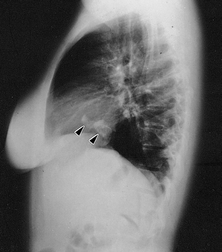
Lateral chest radiograph from a 16-year-old girl with syncope and bacterial endocarditis. The radiograph demonstrates two areas of dense calcification (arrowheads) overlying the posterior aspect of heart. The posterior-anterior (PA) view confirmed location in the heart (not shown). At surgery a calcified myxoma of the right atrium was removed. Image courtesy of Professor Peter Anderson DVM PhD and published with permission © PEIR, University of Alabama at Birmingham, Department of Pathology

| Imaging Technique | Features | Description | Advantages | Limitations |
|---|---|---|---|---|
| Two- or three-dimensional echocardiography |
|
|
|
|
| MRI |
|
|
|
|
| CT |
|
|
|
|
| Angiography |
|
|
|
|
| Chest x-ray |
|
|
|
|
References
- ↑ Cardiac Myxoma. Radiopedia.http://radiopaedia.org/articles/cardiac-myxoma Accessed on November 24, 2015
- ↑ Thyagarajan B, Kumar MP, Patel S, Agrawal A (January 2017). "Extracardiac manifestations of atrial myxomas". J Saudi Heart Assoc. 29 (1): 37–43. doi:10.1016/j.jsha.2016.07.003. PMC 5247297.
- ↑ Reeder GS, Khandheria BK, Seward JB, Tajik AJ (1991). "Transesophageal echocardiography and cardiac masses". Mayo Clin. Proc. 66 (11): 1101–9. PMID 1943240.