Myxoma chest x ray: Difference between revisions
| Line 8: | Line 8: | ||
==Gallery== | ==Gallery== | ||
<div align=" | <div align="center"> | ||
<gallery heights="225" widths="225"> | <gallery heights="225" widths="225"> | ||
Image:Myxoma chest x-ray 1.jpg|Lateral chest radiograph from a 16-year-old girl with syncope and bacterial endocarditis. The radiograph demonstrates two areas of dense calcification (arrowheads) overlying the posterior aspect of heart. The posterior-anterior (PA) view confirmed location in the heart (not shown). At surgery a calcified myxoma of the right atrium was removed. <small>[http://www.peir.net Image courtesy of Professor Peter Anderson DVM PhD and published with permission © PEIR, University of Alabama at Birmingham, Department of Pathology]</small> | Image:Myxoma chest x-ray 1.jpg|Lateral chest radiograph from a 16-year-old girl with syncope and bacterial endocarditis. The radiograph demonstrates two areas of dense calcification (arrowheads) overlying the posterior aspect of heart. The posterior-anterior (PA) view confirmed location in the heart (not shown). At surgery a calcified myxoma of the right atrium was removed. <small>[http://www.peir.net Image courtesy of Professor Peter Anderson DVM PhD and published with permission © PEIR, University of Alabama at Birmingham, Department of Pathology]</small> | ||
Revision as of 17:05, 25 November 2015
|
Myxoma Microchapters |
|
Diagnosis |
|---|
|
Treatment |
|
Case Studies |
|
Myxoma chest x ray On the Web |
|
American Roentgen Ray Society Images of Myxoma chest x ray |
Editor-In-Chief: C. Michael Gibson, M.S., M.D. [1]; Associate Editor-In-Chief: Cafer Zorkun, M.D., Ph.D. [2] Ahmad Al Maradni, M.D. [3]
Overview
There are no specific chest x-ray findings associated with cardiac myxoma, the results can be reported as normal.
Chest x-ray
There are no specific chest x-ray findings associated with cardiac myxoma, the results can be reported as normal. [1] Related imaging findings include cardiomegaly, left atrial enlargement, vascular redistribution, prominent pulmonary trunk, and intracardiac tumoral calcification (rare).
Gallery
-
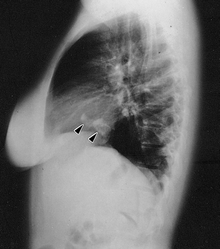
Lateral chest radiograph from a 16-year-old girl with syncope and bacterial endocarditis. The radiograph demonstrates two areas of dense calcification (arrowheads) overlying the posterior aspect of heart. The posterior-anterior (PA) view confirmed location in the heart (not shown). At surgery a calcified myxoma of the right atrium was removed. Image courtesy of Professor Peter Anderson DVM PhD and published with permission © PEIR, University of Alabama at Birmingham, Department of Pathology

References
- ↑ Cardiac Myxoma. Radiopedia.http://radiopaedia.org/articles/cardiac-myxoma Accessed on November 24, 2015