Mumps virus
|
Mumps Microchapters |
|
Diagnosis |
|---|
|
Treatment |
|
Case Studies |
|
Mumps virus On the Web |
|
American Roentgen Ray Society Images of Mumps virus |
| style="background:#Template:Taxobox colour;"|Template:Taxobox name | ||||||||
|---|---|---|---|---|---|---|---|---|
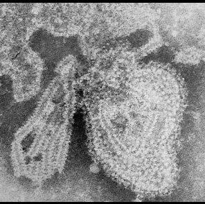
 TEM micrograph of the mumps virus.
| ||||||||
| style="background:#Template:Taxobox colour;" | Virus classification | ||||||||
| ||||||||
| Type species | ||||||||
| Mumps virus |
Editor-In-Chief: C. Michael Gibson, M.S., M.D. [1]; Associate Editor(s)-In-Chief: Lakshmi Gopalakrishnan, M.B.B.S. [2]
Overview
Mumps is caused by a paramyxovirus, and transmission of the virus occurs via respiratory secretions such as infected saliva, air droplets or via direct contact with articles that have been contaminated with infected saliva. The incubation period is usually 18 to 21 days. Infected patients remain contagious from approximately 6 days before the onset of symptoms until about 9 days after the onset of symptoms.
Organism
- Mumps virus (MuV) is an enveloped, non-segmented, negative-sense RNA virus that causes mumps.
- MuV belongs to the genus Rubulavirus and family Paramyxovirus.
Morphology
- The spherical virion is approximately 200nm is diameter and its genome consists of a single RNA strand of 15,384 nucleotides.[1]
- The RNA is encapsidated by nucleoprotein (N protein) forming the ribonucleoprotein (RNP) complex.
Replication Cycle
- MuV binds to host cell sialic acid via haemagglutinin-neuraminidase (HN) and fusion (F) glycoproteins and cause virus-to-cell membrane fusion.
- Replication and transcription is mediated by an RNA polymerase complex composed of large (L) and phospho- (P) proteins.
- Budding is initiated after HN and F glycoproteins are transported through the endoplasmic reticulum and Golgi body to the cell surface.
- Matrix (M) protein localizes the RNP to the area of the host cell expressing HN and F.
Human Pathogen
- Humans are the only natural host of MuV.
- MuV is able to evade an immune response to infection with the following virulence factors:
- Small hydrophobic (SH) protein is presumed to block TNFα-mediated apoptosis.[2]
- Non-structural proteins NS1 and NS2 (V proteins) inhibit IFN production and signaling.[3]
Related Chapters
References
- ↑ Hviid A, Rubin S, Mühlemann K (March 2008). "Mumps". The Lancet. 371 (9616): 932–44. doi:10.1016/S0140-6736(08)60419-5. PMID 18342688.
- ↑ He B, Lin GY, Durbin JE, Durbin RK, Lamb RA (2001). "The SH integral membrane protein of the paramyxovirus simian virus 5 is required to block apoptosis in MDBK cells". J Virol. 75 (9): 4068–79. doi:10.1128/JVI.75.9.4068-4079.2001. PMC 114152. PMID 11287556.
- ↑ Andrejeva J, Childs KS, Young DF, Carlos TS, Stock N, Goodbourn S; et al. (2004). "The V proteins of paramyxoviruses bind the IFN-inducible RNA helicase, mda-5, and inhibit its activation of the IFN-beta promoter". Proc Natl Acad Sci U S A. 101 (49): 17264–9. doi:10.1073/pnas.0407639101. PMC 535396. PMID 15563593.